Cortinarius subfuscoperonatus Y. Li & M.L. Xie, sp. nov. 2020
MycoBank No: 830782
Holotype: China. Gansu Province: Zhangye City, Minle County, Gansu Qilianshan National Nature Reserve, coniferous forest (Picea crassifolia), 38°17'55"N, 100°45'54"E, alt. 2860 m, 9 August 2018, M.L. Xie, HMJAU44444, GenBank No.
(ITS) MK552387.
Morphological description
Pileus 1.6–4.4 cm in diam., hemispherical when young, then low convex, weakly hygrophanous, pale greyish-brown (6C3), sometimes reddish-brown (9E5–9E6) to dark brown (6F6–6F8), surface with greyish-brown fibrillose, margin wavy with age. Lamellae emarginate, medium-spaced, reddish-brown to rusty brown (7D6–7E7), margin even when young, then slightly serrate. Stipe 2.3–7.5 cm long, 0.8–1.3 cm thick at apex, 1.5–2.5 cm thick at base, clavate, white to pale grey (E2), mycelium white at the base. Universal veil greyish-brown (6C2), rich, usually forming an annular band on the middle part and distinct belts or zones lower down. Context white (A1), greyish-brown (7F8) and marbled watery when moist, strongly hygrophanous near pileus and lamellae. Odour somewhat radish-like. Chemical reaction: pileus and context (fresh basidiomata) are dark black brown (8F3) with 10% KOH. Exsiccata brown (6E5) to dark brown (6F5).
Basidiospores 9.5–12.1 × 7.9–9.7 μm, Q = 1.10–1.45, `X = 10.3–11.2 × 8.0– 8.6 μm, `Q = 1.24–1.33 (135 spores, 5 collections), subglobose to broadly ellipsoid, moderately to strongly verrucose, strongly dextrinoid. Basidia 4-spored, clavate, 35–58 × 10–13 μm, thin-walled, hyaline to olivaceous brown in 5% KOH. Lamellar edge fertile, with cylindrical-clavate sterile cells, 13–27 × 6–11 μm, thin-walled, hyaline to slightly olivaceous yellow in 5% KOH. Lamellar trama hyphae regular, pale olivaceous to olivaceous brown in 5% KOH, smooth. Pileipellis: epicutis hyphae cylindrical, 4–12 μm wide, slightly olivaceous brown to olivaceous brown in 5% KOH, some hyphae finely encrusted; hypocutis well developed, hyphae 24–89 × 15–29 μm, sub-cellular to sub-cylindrical, thin-walled, hyaline to slightly olivaceous brown in 5% KOH, smooth. Pileus trama hyphae almost thin-walled, hyaline in 5% KOH, smooth. Clamp connections present.
Habitat: In coniferous forest (Picea crassifolia dominated forest).
Distribution: Gansu Province, China.
GenBank Accession:NO MK552389, MK552390, MK552388, MK552387, MK552391
Notes: Cortinarius subfuscoperonatus corresponds well to the characteristics of section Fuscoperonati, with weak hygrophanous pileus, an annular band on the middle stipe and distinct belts or zones lower down, large spores (> 10 μm long) and grow in coniferous forests. Cortinarius fuscoperonatus was previously placed in section Bovini M.M. Moser (Bidaud et al. 2009; Soop 2014) and Armillati (Brandrud et al. 1992), until Niskanen et al. (2015) placed it in section Fuscoperonati. Cortinarius subfuscoperonatus has remarkably similar morphological characteristics to C. fuscoperonatus, apart from the spores of C. fuscoperonatus being narrower (9.7–11.6 × 6.6–7.7 μm), the pileus being chocolate brown to blackish-brown and being fine fibrous to fine scaly (Schmidt-Stohn et al. 2017). In addition, C. subfuscoperonatus formed a sister relationship with C. fuscoperonatus and was well separated according to the phylogenetic analyses. C. subfuscoperonatus could be considered as the second species in section Fuscoperonati.
Reference: Xie M-L, Wei T-Z, Fu Y-P et al. (2020) Three new species of Cortinarius subgenus Telamonia (Cortinariaceae, Agaricales) from China.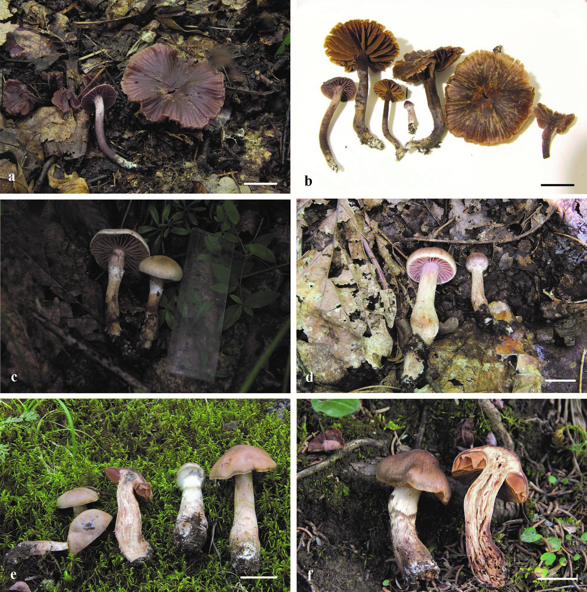
Basidiocarps of three newly-described species. e, f Cortinarius subfuscoperonatus (e HMJAU44444, holotype; f HMJAU44445). Scale bars: 2 cm (a, b, d–f). Photographs by Meng-Le Xie.
Basidiospores of three newly-described species. c Cortinarius subfuscoperonatus (HMJAU44444, holotype). Photographs by Meng-Le Xie.
Margin cells of three newly-described species. c Cortinarius subfuscoperonatus (HMJAU44444, holotype). Photographs by Meng-Le Xie.


