Beltrania dushanensis C.G. Lin, Jian K. Liu & K.D. Hyde, sp. nov. 2020
Index Fungorum Number: IF556728; Facesoffungi number: FoF06220
Holotype: China, Guizhou Province, Qiannan Buyi Miao Autonomous Prefecture, Dushan County, Guizhou Zilinshan National Forest Park (Shengou District), on decaying seeds, 6 July 2018, Chuan-Gen Lin, DS 1-5 (MFLU 19–2252, holotype); ibid. (HKAS 105107, isotype); extype living culture GZCC18–0020.
Morphological description
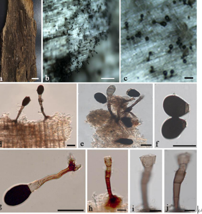
Saprobic on decaying seed. Sexual morph: Undetermined. Asexual morph: Colonies on plant substrate effuse, dark brown, velutinous. Mycelium mostly immersed in the substratum. Setae numerous, erect, straight or slightly flexuous, unbranched, thick-walled, smooth, pale brown to dark brown, 180–260 μm long, 4–8 μm wide at the base, tapering to a pointed apex, arising from a dark brown, swollen, radially lobed basal cell, 10–17 μm diameter Conidiophores macronematous, single or in small groups, straight or flexuous, septate, smooth, thick-walled, cylindrical or clavate, pale brown, 10–45 μm long, 4–7 μm wide, arising from basal cells of setae or from separate dark brown, swollen, radially lobed cells, 8–18.5 μm diameter, often reduced to conidiogenous cells. Conidiogenous cells polyblastic, integrated, terminal, sympodial, denticulate, cylindrical, clavate, pale brown, smooth, 6.5–24 µm long, 4.3–8.5 µm wide. Separating cells obovoid, ellipsoidal, smooth, thin-walled, hyaline, 7.9–12.7 µm (x̄ = 10.5 μm, n = 23) long, 4.4–5.8 µm (x̄ = 5.1 μm, n = 23) wide in the broadest part. Conidia arise directly from conidiogenous cells or from separating cells, acrogenous, simple, smooth, dry, straight, biconic, rostrate, appendiculate, pale brown with a subhyaline equatorial transverse band, 21.4–27.9 µm (x̄ = 24.3 μm, n = 30) long, 8.9–12.6 µm (x̄ = 10.4 μm, n = 30) wide in the broadest part; appendage 6.3–12.1 µm long, 0.7–1.6 µm wide at the base, tapering to a pointed apex.
Habitat: on decaying seeds
Distribution: China
GenBank Accession: ITS MN252875 ;LSU MN252882
Notes: Beltrania dushanensis is most similar to B. rhombica Penz. in having biconic, symmetrical, light brown conidia with a V-shaped proximal end, but differs by its short conidiophores which are often reduced to conidiogenous cells (Pirozynski 1963, Morelet 2001). In addition, B. dushanensis and B. rhombica can be recognized as phylogenetically distinct lineages (Fig. 154). Phylogenetically, B. dushanensis shows a close relationship with B. krabiensis Tibpromma & K.D. Hyde (Fig. 154) since they clustered and formed a monophyletic group, however, they can be recognized as distinct lineages. Beltrania dushanensis differs from B. krabiensis by its shorter conidiophores and larger conidia (Tibpromma et al. 2018b).
Reference: Hyde KD, de Silva NI, Jeewon R et al. 2020 – AJOM new records and collections of fungi: 1–100. Asian Journal of Mycology 3(1), 22–294, Doi 10.5943/ajom/3/1/3 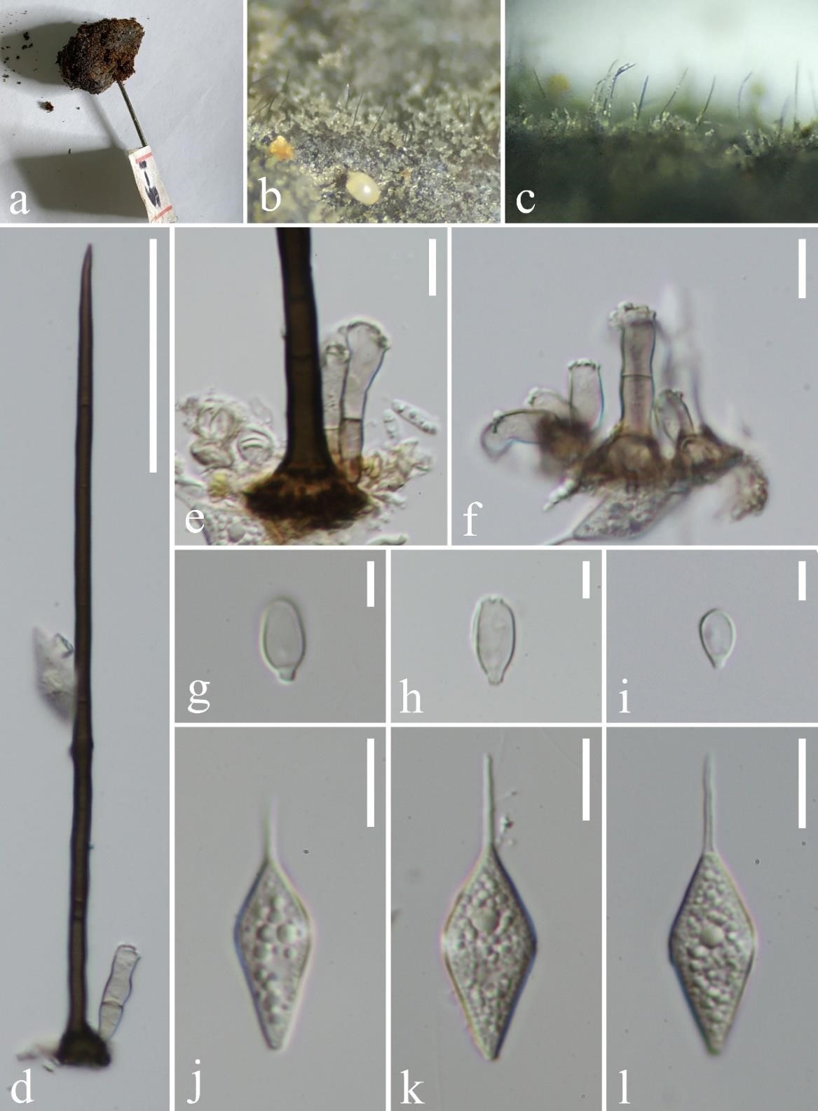
Beltrania dushanensis (MFLU 19–2252, holotype). a Host material. b, c Conidiophores on leaf surface. d Setae, conidiophores and conidiogenous cell. e, f Conidiogenous cells. g–i Separating cells. j–l Conidia. Scale bars: d = 50 μm, e, f, j–l = 10 μm, g–i = 5 μm.


