Cytospora pingbianensis Q.J. Shang, K.D. Hyde & J.K. Liu, sp. nov. 2020
Fungorum number: IF 555514; Facesoffungi number: FoF 05107
Holotype: CHINA, Yunnan Province, Pingbian, on a dead branch of undetermined wood, 26 September 2017, Qiuju Shang, PB45 (KUN-HKAS 102161, holotype; MFLU 18-1389, isotype), ex-type living culture, MFLUCC 18-1204.
Morphological description
Saprobic on the bark. Sexual morph Stromata 880–1524 µm wide, with the poorly developed interior, solitary to gregarious, immersed, becoming raised to erumpent by the ostiolar canal, dark brown to black, glabrous, circular in shape, arranged with conspicuous, clustered, roundish to cylindrical prominent ostioles. Ascomata (excluding necks) 142–248 μm high, 113–245 µm diam. (x = 195 × 180 μm, n = 50), perithecial, immersed in a stroma, brown to dark brown, globose to subglobose, glabrous, individual ostiole with the neck. Ostiolar canal 185–722 μm high, 27–66 μm diam. (x = 453 × 47 μm, n = 10), cylindrical, sulcate, periphysate. Peridium 14–24 μm wide, composed of two section layers, outer section comprising 3–5 layers, of relatively small, brown to dark brown, thick–walled cells, arranged in textura angularis, the inner part comprising 3–5 layers of hyaline cells of textura angularis. Hamathecium comprising only asci. Asci (25–)27–30(–33) × (3.5–)4–5(–6) μm ( x = 28 × 4.7 μm, n = 70), 8-spored, unitunicate, clavate, sessile, apically rounded to truncate, with a J- apical ring. Ascospores (4.6–)5.8–6.7(–7.5) × (1–)1.5–2(–2.5) μm (x = 6.2 × 1.7 μm, n = 210), overlapping 1–2-seriate, hyaline, allantoid, aseptate, smooth-walled. Asexual morph Undetermined. Culture characteristics – Ascospores germinating on PDA within 12 hrs. Germ tubes produced from all sides. Colonies on PDA reaching 2.5–5.5 cm diam. after 5 days at room temperature, colonies circular to irregular, medium dense, flat or effuse, slightly raised, with edge fimbriate, fluffy to fairy fluffy, initially white to yellow from above, yellow from below; After 10 days, yellow to brown from above, brown to dark brown from below; not producing pigments in agar.
Habitat: on a dead branch of undetermined wood
Distribution: Yunnan, China
GenBank Accession: ITS MK912135; LSU MK571763; ACT MN685817; RPB2 MN685826
Notes: The phylogenetic result (Fig. 1) shows that our strain of Cytopora pingbianensis (MFLUCC 18-1204) forms a distinct lineage and is close to Cytopora platycladi (CFCC 50504, CFCC 50505) with the high support (100% ML, 99% MP and 1 PP). These taxa form a sister clade to Cytopora lumnitzericola with moderate support (89% ML, 75% MP and 0.99 PP). Cytospora platycladi and C. lumnitzericola were reported as asexual morph taxa associated with canker disease (Norphanphoun et al. 2018, Fan et al. 2020), while our taxon is only reported as a sexual morph. In this study, C. pingbianensis can be recognized as a phylogenetically distinct species (Fig. 1), and it is introduced as new species with detailed description and illustration of the sexual morph. Culture characteristics – Ascospores germinating on PDA within 12 hrs. Germ tubes produced from all sides. Colonies on PDA reaching 2.5–5.5 cm diam. after 5 days at room temperature, colonies circular to irregular, medium dense, flat or effuse, slightly raised, with edge fimbriate, fluffy to fairy fluffy, initially white to yellow from above, yellow from below; After 10 days, yellow to brown from above, brown to dark brown from below; not producing pigments in agar.
Reference: Shang QJ, Hyde KD, Camporesi E et al. 2020 – Additions to the genus Cytospora with sexual morph in Cytosporaceae. Mycosphere 11(1), 189-224, Doi 10.5943/mycosphere/11/1/2 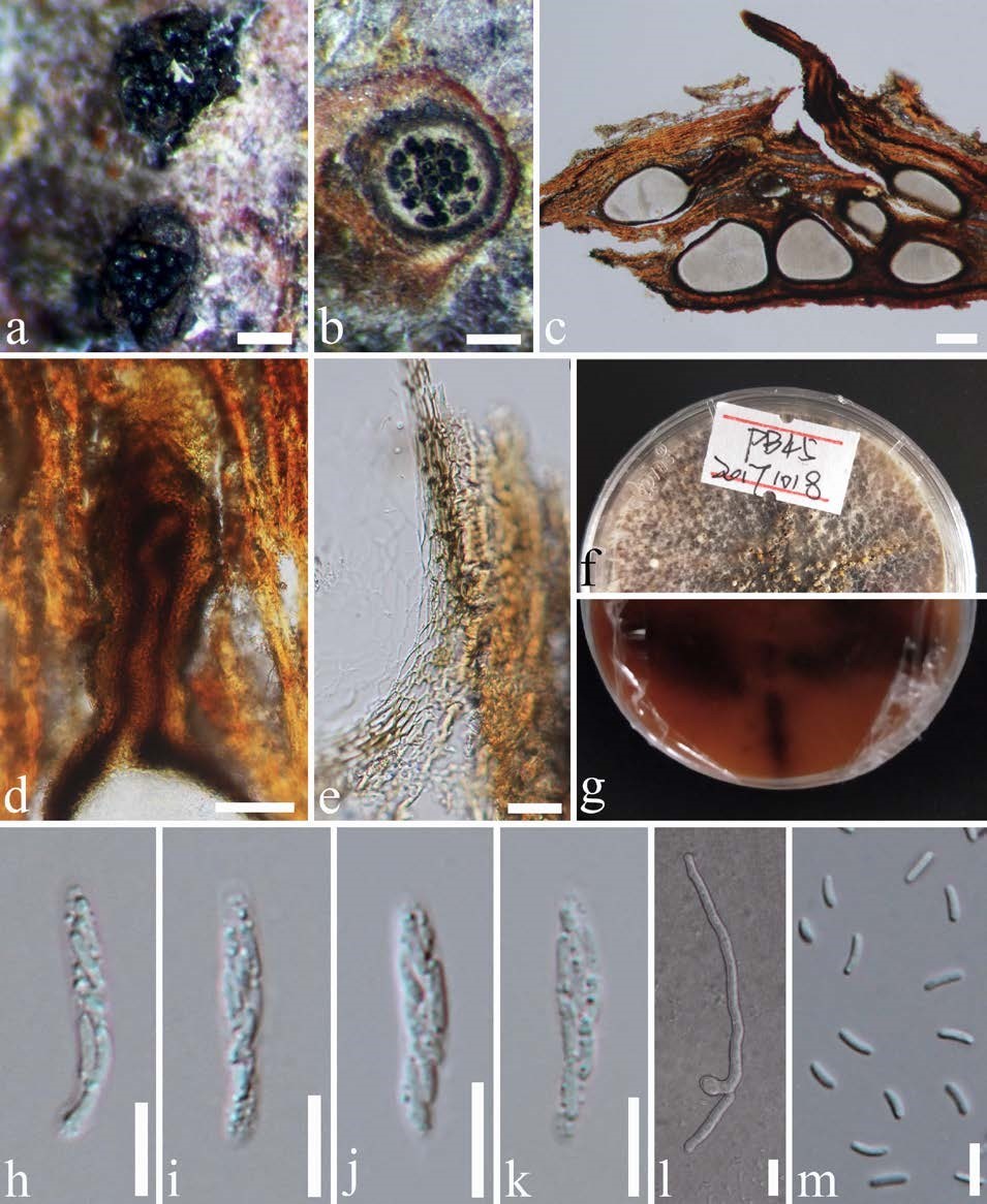
Cytospora pingbianensis (KUN-HKAS 102161, holotype). a Stromata. b Cross section through stroma. c Vertical section through stroma. d Ostiolar canal. e Peridium. f, g Culture characteristic on PDA (f = colony from above, g = colony from below). h–k Asci. l Germinating ascospore. m Ascospores. Scale bars: a, b = 200 µm, c = 100 µm, d = 50 µm, e, l = 20 µm, h–k, m = 10 µm.


