 83
83
Alternaria vulgarae L. He & J.X. Deng, sp. nov.2021
MycoBank No: 838892
Holotype: China, Hubei Province, Yichang city, Badong county on infected leaves of Foeniculum vulgare. 19 July, 2016, J.X Deng, (YZU-H-0040, holotype), ex-type cul ture YZU 161234.
Morphological description
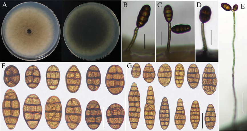
Sexual morph: Colonies on PDA (Fig. 3A) hazel in center and vinaceous buff at the edge, greenish black to mouse gray in reverse, surface velvety or floccose, 79‒82 mm in diam.; On PCA, conidiophores straight or curved, 12–80 × 4–6 μm, 1‒4 transverse septa; conidia solitary arising from the apex or near the apex of the conidiophores or terminal hyphae, rare from lateral of wire-like hyphae, ovoid, short-ovoid or ellip soid, 25–50 (–70) × 16–27 μm, with 1–5 transverse septa and 1‒4 longitudinal septa (Fig. 3B, C, F); On V8A, conidiophores 24–93 × 4–7 μm, 1‒4 transverse septa, wire like hyphae up to 200–400 × 4–6 μm; conidia short-ovoid, ovoid, ellipsoid or long ellipsoid, 24–55 (–77) × 13–26 μm, 1‒8 transverse septa, 1‒4 longitudinal or oblique between septa (Fig. 3D, E, G). There was no secondary conidium production observed on PCA and V8A medium.
Asexual morph: Undetermined
Culture characteristics:
Habitat:
Distribution: China
GenBank Accession:
ITS MW541936; GAPDH MW579308; TEF1 MW579310; RPB2 MW579312
ITS MW541937; GAPDH MW579309; TEF1 MW579311; RPB2 MW579313
Notes: Phylogenetic analysis based on combining four gene fragments indicated that Alternaria vulgarae fell in an individual branch in section Radicina of Alternaria and displayed a close relationship with A. petroselini and A. selini with high supported values (Fig. 1). Morphologically, A. vulgarae could be easily distinguished from A. petroselini and A. selini by their sporulation and length of conidiophores. Conidia of A. petroselini were solitary or cluster a small clump with 2‒4 spores near the tips or lateral of con idiophores. Occasionally, the secondary conidium could be observed. Meanwhile, the single conidium or conidial chains (1–3) of A. selini grew from numerous lateral conidi ophores, which produced from wire-like hyphae (Simmons 2007). Differently, conidia of A. vulgarae were erected from apex of conidiophores or terminal hyphae. There were no small conidial clumps and secondary conidium formed (Fig. 3B, C, D, E). Moreo ver, the conidiophores of A. vulgarae (12–80 × 4–6 μm) was longer than A. petroselini (30–60 × 5–6.5 μm) and shorter than A. selini (200–400 × 4–6 μm) (Simmons 2007). Besides, A. vulgarae differed from A. petroselini in conidial shape. Conidium popula tions of A. petroselini were dominated by shot-ovoid to subsphaerical spores though, the shapes of A. vulgarae were mainly ovoid, ellipsoid or long-ellipsoid (Simmons 2007).
Reference: [1] He, L. , Cheng, H. , Zhao, L. , Htun, A. A. , & Li, Q. L. . (2021). Morphological and molecular identification of two new alternaria species (ascomycota, pleosporaceae) in section radicina from china. MycoKeys, 78, 187-198.
Morphological characteristics of Alternaria vulgarae (strain: YZU 161234). Colony on PDA for 7 days at 25 °C (A); Sporulation patterns on PCA and V8A (B–E: B, C from V8A D, E from PCA); Conidia from PCA and V8A (F–G). Scale bars: 25 μm (B, C, D, F, G); 50 μm (E).

