Gymnosporangium lachrymiforme P. Zhao & L. Cai 2020
MycoBank MB832743
Holotype: China, Guizhou, Guiyang City, 0, I on Malus sp., 14 May 2015, P. Zhao (holotype HMAS248123). ITS and sequences GenBank MN605716 and MN605794.
Morphological description
Spermogonia, uredinia and telia not found. Aecia foliicolous, hypo- phyllous, roestelioid; peridium balanoid, 3.0–8.5 mm high, rupture and becoming lacerate at side, peridial cells oblong, 47–85 × 24–33 μm, outer walls smooth, inner walls and side walls densely verrucose with small papillae; aeciospores globoid or broadly ellipsoid, 15–29 × 14–24 μm, walls slightly brown, 2 μm thick; germ pores scattered, 3–7.
Habitat: On Malus sp.
Distribution: In China.
GenBank Accession:
Notes: This rust was found on one unidentified Malus species in China, and it was characterised by its balanoid aecia with linear and relatively shorter peridia. This species resembles G. sabinae in its balanoid aecia, but the length of peridia, and dimensions of the peridial cells clearly differentiate the two species. Among other Gymnosporangium species reported on Malus, the aecial stage of this species only resembles G. hemisphericum in the dimension and shape of peridial cells and aeciospores (Hiratsuka et al. 1992). However, our new species differs in the balanoid aecia with relatively long peridia. Based on these morphological and molecular differences, we proposed it as a new species.
Reference: P. Zhao1, X.H. Qi 2, P.W. Crous3,4 et al.
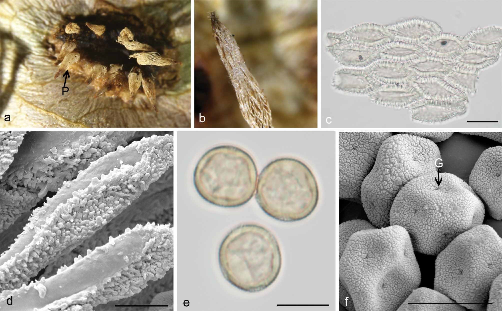
Morphology of G. lachrymiforme. a. Aecia with balanoid peridia (P) on the hypophyllous leaf surface; b. peridium with cornuted apex; c. oblong peridial cells observed by LM; d. ultrastructure of peridial cells observed by SEM; d. globoid or ellipsoid aeciospores with scattered germ pores (G) observed by LM; e. ultrastructure of aeciospores observed by SEM. — Scale bars: c–f = 20 µm.