Auxarthron guangxiense Z.F. Zhang & L. Cai, sp. nov. 2020
Index Fungorum number: 556413;Facesoffungi number: FoF 08432
Holotype: CHINA, Guangxi, Guilin, E’gu Cave, N 24.942°, E 110.511°, on soil, May 2016, Z.F. Zhang, HMAS 247993 (holotype designated here), ex-type living culture CGMCC3.19634 = LC12464; ibid., LC12465.
Morphological description
Hyphae hyaline, septate, branched, smooth, 1.5–2.5 μm diam. Sexual morph Ascomata abundant, solitary or in clusters, subglobose to globose, white at first, becoming orange-brown at maturity, 250–380 μm diam. Peridial hyphae rough, thick-walled, septate, pale brown, branched and anastomosed to form a reticuloperidium, terminated by spine-like or blunt prominences, sometimes dichotomously branched, 1.5–2.5 μm diam, appendages not observed. Asci 8-spored, pyriform, subglobose or globose, hyaline, 8.5–12.0 × 6.5–9.0 µm. Ascospores oblate, smooth, hyaline, 2.5–3.5 µm ( x̄ ± SD = 3.1 ± 0.22 µm, n = 40). Asexual morph not observed. Culture characteristics—Colonies on PDA attaining 26–31 mm diam. after 4 weeks, flat, margin crenate, cottony, cream-white (2A1) to yellow (2A3) at fruiting region, floralwhite at aging region. Reverse pale yellow (1A2) to goldenrod (2A3) at margin, dark brown (4D8) at center. Colonies on OA attaining 32–40 mm diam. after 4 weeks, flat, annular, cottony at middle, white to pale yellow (2A3) from margin to center. Reverse pale yellow (2A3). Colonies on SNA attaining 28–32 mm diam. after 4 weeks, flat, white to pale yellow (1B3), aerial mycelia sparse, with ascomata scattered. Reverse white to pale yellow (1B3). Sporulation within 3 weeks on SNA.
Habitat: on soil.
Distribution: Guangxi, Guilin, E’gu Cave, China.
GenBank Accession:
Notes: Phylogenetically, Auxarthron guangxiense is close to A. pseudauxarthron G.F. Orr & Kuehn (Fig. 15), but differs in the absence of ascomatal appendages.
Reference: Zhi‑Feng Zhang ,Shi‑Yue Zhou ,Lily Eurwilaichitret al.
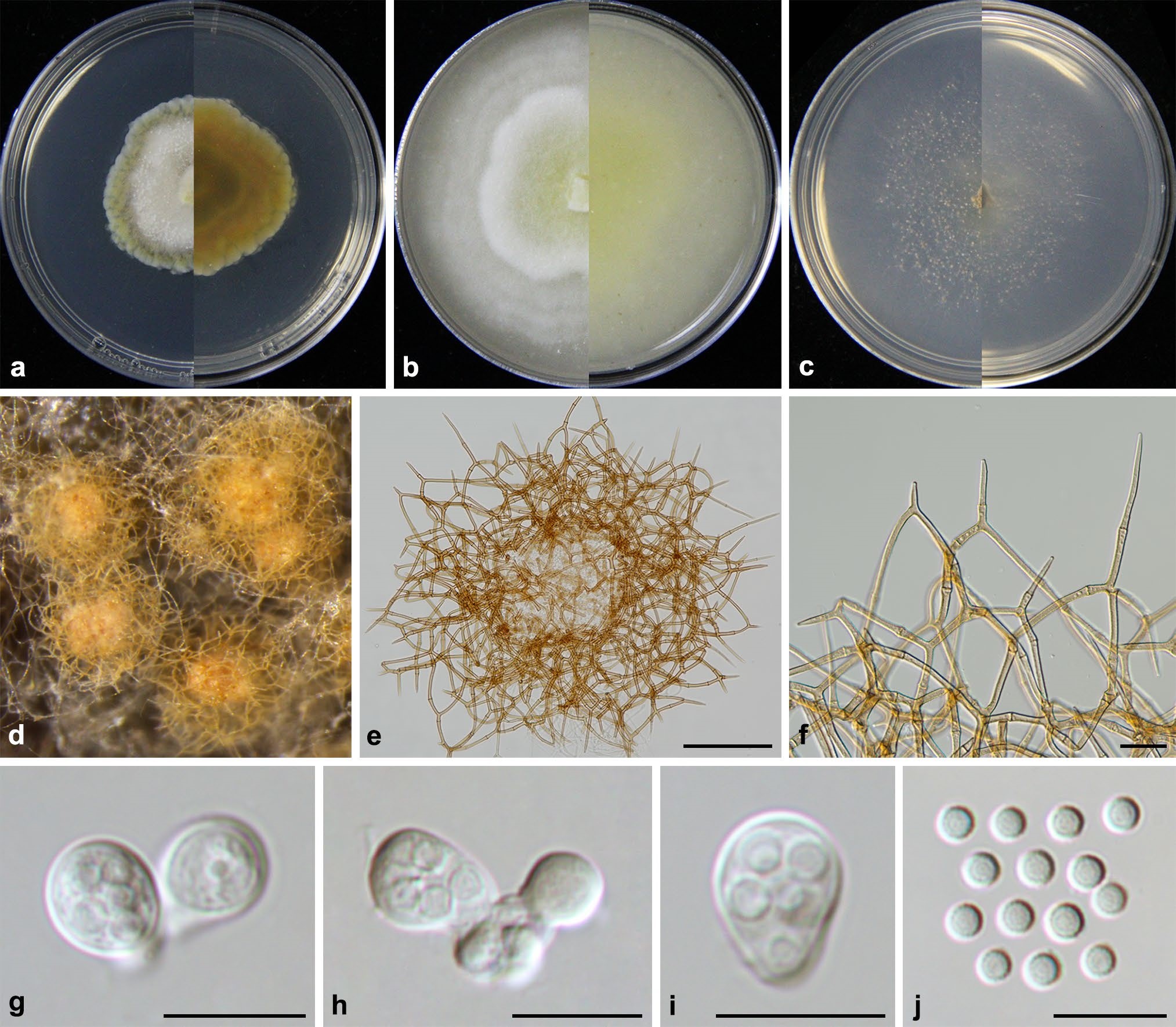
Auxarthron guangxiense (from ex-holotype CGMCC3.19634). a–c Upper and reverse views of cultures on PDA, OA and SNA 4 weeks after inoculation; d ascomata; e, f peridial hyphae; g–i asci; j ascospores. Scale bars: e 50 μm; f 20 μm; g–j 10 μm