Trichoderma lentinulae Jing Z. Sun & X.Z. Liu, sp. nov.2020
MycoBank No: 833233
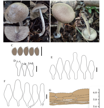
Holotype China. Haidian District, Beijing, 39°57'40"N, 116°19'40"E, ca. 27 m elev., from a fruiting body and mushroom spawn of Lentinula edodes, 19 Oct 2018, Jing Z. Sun (HMAS 248256, holotype), ex-type culture CGMCC 3.19847.
Morphological description
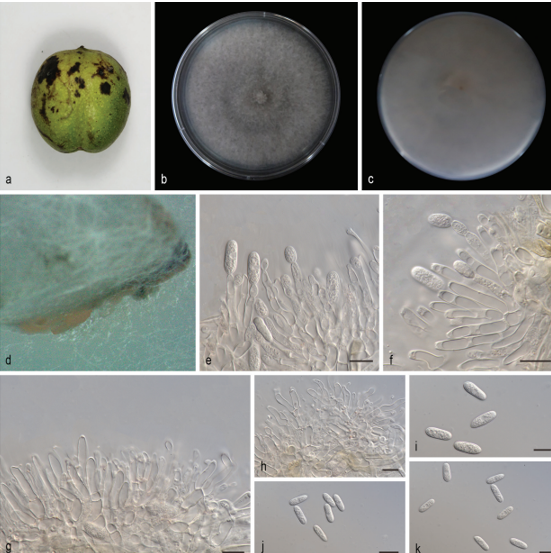
On CMD after 72 h, colony radius 57–58 mm at 25 °C, covering the plate at 30 °C, 4–5 mm at 35 °C. Colony hyaline, weak, indistinctly radial. Aerial hyphae short, inconspicuous. No diffusing pigment noted, odor indistinct (Fig. 2B). Conidial production noted after 3 days, scant, effuse in aerial hyphae, becoming bluegreen after 7 days. Chlamydospores not observed.
On PDA after 72 h, colony radius 45–46 mm at 25 °C, mycelium covering the plate at 30 °C, 11–12 mm at 35 °C. Colony white to yellowish-white, regularly circular, indistinctly zonate; mycelium dense and radial. No diffusing pigment, not distinct odor (Fig. 2A). Conidial production noted after 3 days, starting around the original inoculum, effuse in the aerial hyphae, first white, turning green after 3 d. Chlamydospores unobserved.
On SNA after 72 h, colony radius 51–52 mm at 25 °C, 52–53 mm at 30 °C, 4–5 mm at 35 °C. Colony hyaline, indistinctly zonate; mycelium loose, especially at the margin. Aerial hyphae loose. No diffusing pigment, not distinct odor (Fig. 2C). Conidial production noted after 2 days, starting around the inoculum, effuse in the aerial hyphae. Small pustules formed around the inoculum, first white, turning green after 3 d, with hairs protruding beyond the surface. Conidiophores pyramidal with opposing branches, less frequently solitary, closely-spaced branches, each branch, and the main axis terminating in 2–5 cruciately to nearly verticillately disposed phialides (Fig. 2F, H, I). Phialides ampulliform, typically strongly constricted below the tip, less frequently lageniform and then usually apex and inequilateral to strongly curved, hyaline, (3.5– )4.0–6.0(–6.5) × (2.0–)2.5–3.0(–3.5) µm ‒(x = 4.5 × 3.0 µm, n = 30), length/width ratio (1.5–)2.0–3.0(–5.0) (‒x = 2.0, n = 30), base 1.0–2.5 µm ‒(x = 1.5 µm)(Fig. 2G, H, I). Conidia ovoid to globose, smooth, hyaline when young, becoming green to dark green with age, (2.0–)2.5–3.0(–3.5) × (1.5–)2.0–2.5(–3.0) µm (‒x = 2.5 × 2.2 µm, n = 50), length/width ratio (1.0–)1.1–1.4 (–1.5) ‒(x = 1.2, n = 50) (Fig. 2J). Chlamydospores common, apex or intercalary, ellipsoid or subglobose, (3.5–)5.0–6.5(–7.0) × (3.0–)4.0–5.0(–6.0) µm ‒(x = 5.5 × 4.5 µm, n = 30), length/width ratio (1.0–)1.2–1.5 (–1.7) ‒(x = 1.2, n = 30) (Fig. 2K–M).
Habitat: fruiting body and mushroom spawn of Lentinula edodes
Distribution: China.
GenBank Accession: ITS MN594469、MN594470、MN594471、MN594478、MN594479 RPB2MN605867、 MN605868、MN605869、MN605876、MN605877 TEF1-a MN605878、MN605879、MN605880、MN605887、MN605888
Notes: The species is characterized by tree-like conidiophores, phialides verticillate or in whorls of 3–4, spindle-like to fusiform phialides (4.0–6.0 × 2.5–3.0 µm) and ovoid to subglobose conidia. Differs from T. lixii by shorter and wider phialides and smaller conidia. Differs from Trichoderma xixiacum by compact, relatively smaller phialides, and the pustules not forming distinctly zonate of pustules on SNA.
Reference: Gu X, Wang R, Sun Q, Wu B, Sun J-Z (2020) Four new species of Trichoderma in the Harzianum clade from northern China. MycoKeys 73: 109–132. https://doi.org/10.3897/mycokeys.73.51424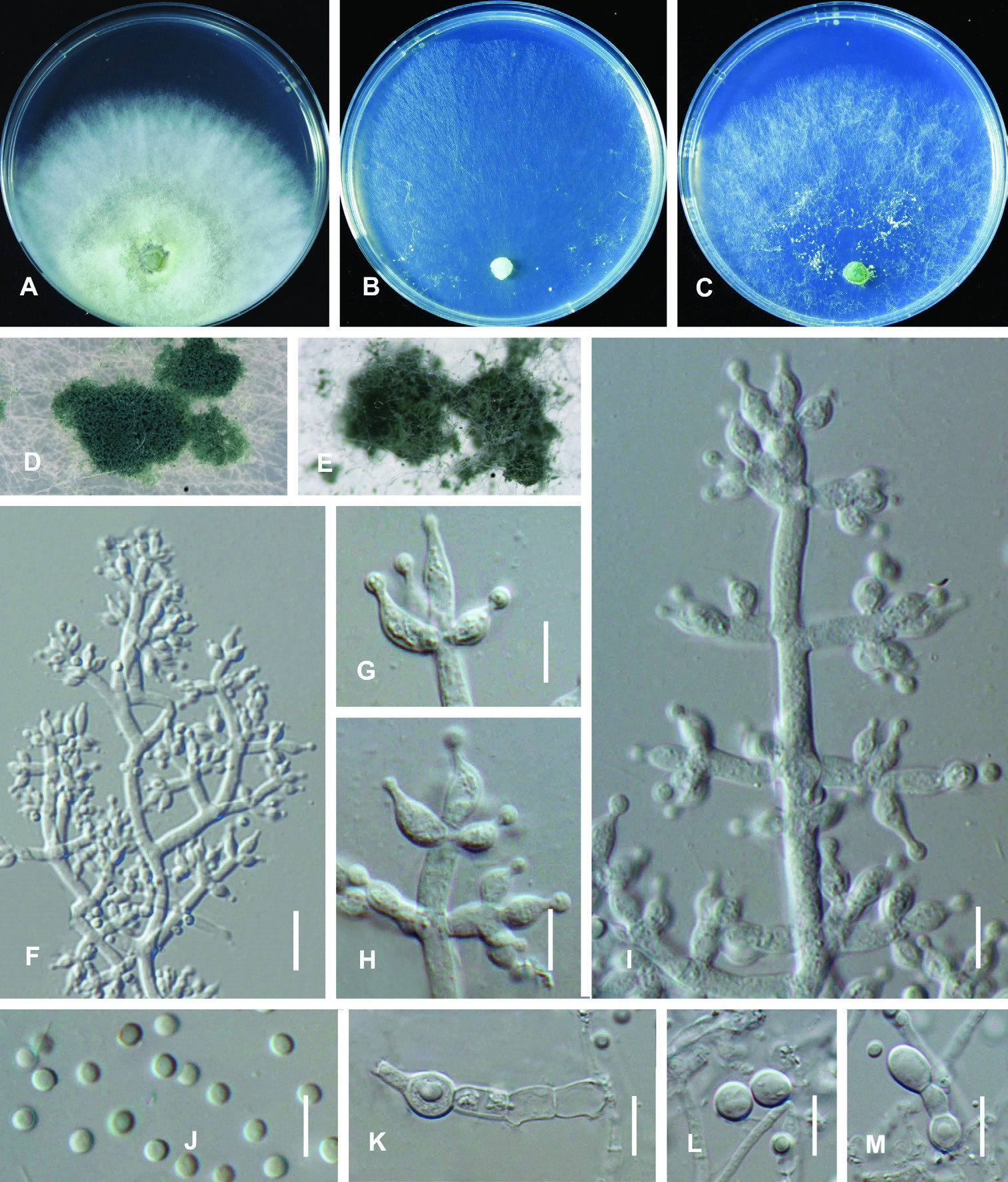
Figure 2. Trichoderma lentinulae (CGMCC 3.19847). Cultures at 25 °C after 3 days (A on PDA B on CMD C on SNA) D conidiation pustules on CMD after 10 days E conidiation pustules on CMD after 10 d F conidiophores G–I Conidiophores and phialides J conidia K–M chlamydospores. Scale bars: 25 µm (F); 10 µm (G–M).