因为
值得
专业,
信赖
 299
299
Amphisphaeria micheliae Samarak., Jian K. Liu & K.D. Hyde 2020
MycoBank MB836112; Facesoffungi number: FoF08752
Holotype: China, Sichuan Province, Chengdu, University of Electronic Science and
Technology of China (UESTC) campus, on a dead branch of Michelia alba (Magnoliaceae), 30 September 2019, M.C. Samarakoon, SAMC244 (HKAS 107012, holotype; MFLU 20-0503, isotype); ex-type living culture MFLUCC 20-0121.
Morphological description
Sexual morph: Ascomata: 180–210µmhigh×225–370µm diameter (M = 190 × 300 µm, n = 8), immersed, visible as black spots in light coloured area on the host, solitary, scattered, subglobose to oblate, papillate ostiole 60–76 µm high × 34–50 µm diam. (M = 68 × 42 µm, n = 8), centric. Periphyses: 1–2 µm wide (M = 1.5 µm, n = 20), hyaline, short. Peridium: two-layered; outer layer: 6.5–11.5 µm (M = 8.5 µm, n =10), dense, reddish brown cells of textura angularis 6.5–12.5 × 1–2.5 µm (M = 9 × 1.5 µm, n = 15), thick-walled. Inner layer: 4–8 µm (M = 6 µm, n =10), loosely arranged, hyaline cells of textura angularis 10.5–17.5 × 2.5–4 µm (M = 14.5 × 3 µm, n = 15), thin-walled, loosely arranged. Paraphyses: 3.5–4.5 µm wide (M = 4 µm, n = 20), hyaline, highly delicate, cellular, constricted septate, guttulate, embedded in a gelatinous matrix. Asci: 92–135 × 7–10.5 µm (M = 115 × 8.5 µm, n = 25), 8-spored, unitunicate, cylindrical, thin-walled, short-pedunculate, apically rounded, with a J+, discoid apical ring. Ascospores: 15.5–21 × 6–7.5 µm (M = 18 × 6.5 µm, n = 40), l/w 2.7, uniseriate, oblong or narrowly fusiform, first hyaline, guttulate, turning yellow to yellow-brown, 1-septate, slightly constricted at septum, straight to slightly curved, smooth-walled. Asexual morph: Undetermined.Culture characteristics: colonies on PDA, reaching 20–22 mm diameter after one week at 25 ◦C; colonies are flat, circular, and dense, with a smooth surface, entire margin, concentrically zonate, white to light brown, media becoming pale brown; reverse yellowish brown at center and light brown edges.
Habitat: dead branch of Michelia alba.
Distribution:China.
GenBank Accession:LSU:MT756619;ITS:MT756625;PRB2:MT789854;TUB2: MT774370
Notes: Two of our collections have solitary, immersed ascomata with two-layered peridium, unitunicate asci with J+, discoid apical ring, and brown ascospores. This collection also has 1-septate ascospores similar to Amphisphaeria unisepta and A. camelliae. Compared to those two similar species, this collection has subglobose to oblate ascomata and oblong or narrowly fusiform ascospores. The phylogenetic analyses show MFLUCC 20-0121 and HKAS 107012 are closely related to A. sambuci, isolated from partly decorticated branches of Sambucus nigra. Amphisphaeria sambuci is different from our new collection in having large, depressed globose ascomata and oblong-ellipsoid, straight, rarely curved, 2–4(–6)-distoseptate ascospores with a thick mucilaginous sheath. Based on the morphology and phylogeny, here we introduce it as the novel species A. micheliae.
Reference: Milan C. Samarakoon 1,2,3,4,5, Sajeewa S. N. Maharachchikumbura , Jian-Kui (Jack) Liu 4 et al.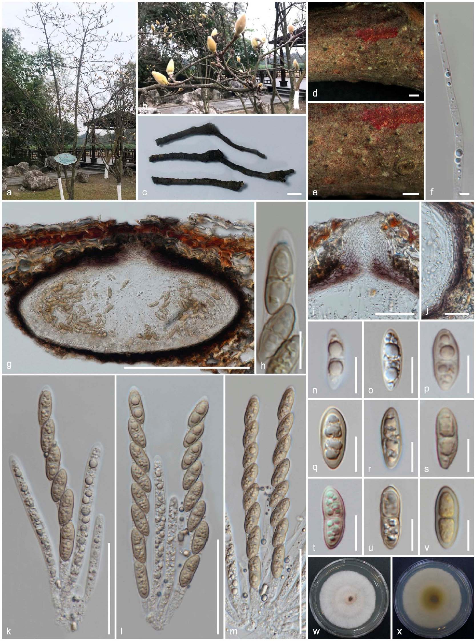
Sexual morph of Amphisphaeria micheliae (HKAS 107012, holotype). (a,b) Host Michelia alba; (c) substrate; (d,e) ascomata on the substrate; (f) paraphyses; (g) vertical section of ascoma; (h) apical ring bluing in Melzer’s reagent; (i) ostiole; (j) peridium; (k–m) asci; (n–v) ascospores (v in Melzer’s reagent); (w) upper view, (x) reverse view of the 2 weeks old colony on PDA. Scale bars are set at (c) 1 cm; (d,e) 500 µm; (g) 200 µm; (i,k–m) 50 µm; (j) 20 µm; (h,n–v) 10 µm.


长按屏幕识别二维码
打开手机扫描二维码