Neomyrmecridium guizhouense N.G. Liu, K.D. Hyde & J.K. Liu 2020
Index Fungorum number: IF557234; Facesoffungi number: FoF 06704
Holotype: CHINA, Guizhou Province, Dushan, on decaying wood in a bank of a small freshwater, 6 July 2018, N.G. Liu, DS002 (GZAAS 20-0001, holotype), extype living culture, GZCC 20-0008.
Morphological description
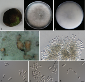
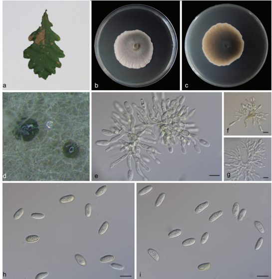
Sexual morph Undetermined. Asexual morph Hyphomycetous. Colonies on natural substrate effuse, greyish white, velvety. Mycelium immersed, composed of brown, branched, septate hyphae. Conidiophores 75–140 × 2–4.5 μm ( x̄ = 98.4 × 3.3 μm, n = 15), macronematous, mononematous, solitary, erect, straight or slightly flexuous, subcylindrical, unbranched, septate, medium brown at base, paler towards apex, smooth, thick-walled. Conidiogenous cells 2.2–4.3 μm ( x̄ = 3.2 μm) wide, polyblastic, terminal, integrated, subcylindrical, subhyline, finely verrucose, with several denticles at apex, not thickened nor darkened. Conidia 8.9–12.7 × 2.8–4.8 μm ( x̄ = 10.4 × 4.1 μm, n = 30), solitary, subhyaline to pale brown, (2–)3-septate, rarely constricted at septum, smooth-walled, guttulate, fusoid-ellipsoid, apex obtuse or tapering, with a subtruncate hilum at base.Culture characteristics: Conidia germinating on water agar within 24 h. Germ tubes produced from one or both ends. Mycelia superficial, circular, with entire edge, mycelia dense at centre, sparse towards circumference, yellowish white to yellow from above, yellow at centre, paler towards circumference from below.
Habitat: decaying wood.
Distribution:China.
GenBank Accession: ITS:MT002305;LSU:MT002307;SSU:MT002308;RPB2:MT023016; TEF1-α:MT023013.
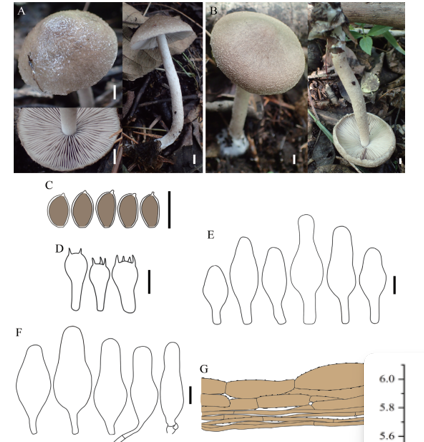
Notes: Neomyrmecridium guizhouense resembles N. asiaticum in having pale brown, (2–)3-septate conidia. However, N. guizhouense has smaller conidia (8.9–12.7 × 2.8–4.8 versus 15–16 × 4.5 μm) and longer conidiophores (75–140 versus 50–100 μm) than those of N. asiaticum. The conidia of N. guizhouense are also smaller than those of N. septatum (8.9–12.7 × 2.8–4.8 versus 14–16 × 4 μm). Phylogenetic analyses of a combined LSU, SSU, ITS and RPB2 sequence dataset showed that N. guizhouense forms a sister lineage with N. septatum with high support (100% ML, 1.00 BYPP; Fig. 120). We did not observe the generic character that upper two-thirds of conidia are encased in mucoid sheath, but phylogenetic analyses confirmed that our taxon belongs to Neomyrmecridium (Fig. 120). Therefore, N. guizhouense is introduced as a new species.
Reference: Kevin D. Hyde1,5,8,22 · Yang Dong2,3 · Rungtiwa Phookamsak1,5,6,7 et al.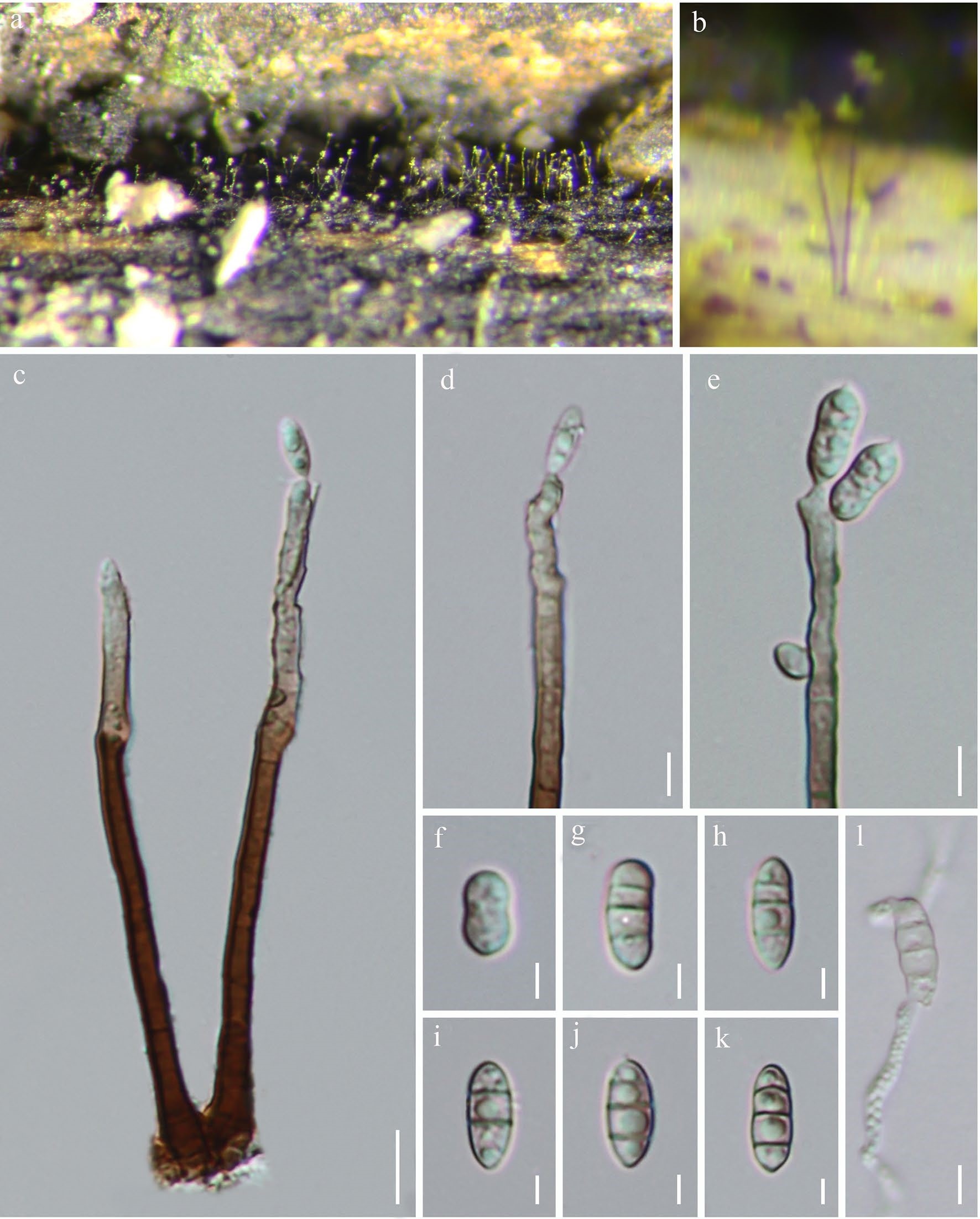
Neomyrmecridium guizhouense (GZAAS 20-0001, holotype). a, b Colonies in natural substrates. c Conidiophores and conidia. d, e Conidiogenous cells and young conidia. f–k Conidia. l Germinated conidium. Scale bars: c = 10 μm, d, e = 5 μm, f–k = 3 μm, l = 5 μm