Conidiobolus taihushanensis B. Huang & Y. Nie, sp. nov. 2020
MycoBank No: 835124; Facesoffungi: FoF 08143
Holotype: China, Anhui: Ma’anshan City, Hanshan County, Taihushan National Forest Park, 31°30'53"N, 118°2'49"E, from plant detritus, 12 Jan 2019, Y. Nie and Y. Cai (holotype HMAS 248724, ex-holotype culture CGMCC 3.16016 = RCEF 6559, GenBank: nucLSU = MT250086; TEF1 = MT274290; mtSSU = MT250088).
Morphological description
Colonies on PDA at 21 °C after 3 d, white, reaching ca. 11–14 mm in diameter. Mycelia colourless, straight and unbranched when young, 8.5–12 μm wide; distended and non-contiguously segmented when old, 10–20 μm wide. Primary conidiophores arising from the older mycelia without an upward widening near the tip, colourless, 44–180 × 7–13 μm, usually unbranched and often producing a single globose primary conidium, at the initial growth stage 2–5 short branches bearing a primary conidium each. Primary conidia forcibly discharged, mostly subglobose, 27–42 × 19–32 μm, with tapering and pointed papilla, 4–10 × 8–12 μm. Secondary conidia arising from primary conidia, similar to, but smaller than the primary ones, forcibly discharged. On 2% water agar, microconidia not observed. Zygospores usually formed between adjacent segments of the same hypha after 5 d, 34–48 × 23–40 μm, with a 2–4 μm thick wall, ellipsoid and rich in content when young, smooth, mostly globose, subglobose to ovate when mature.
Habitat: from plant detritus
Distribution: Anhui Province, China.
GenBank Accession: nucLSU MT250086; EF-1α MT274290; mtSSU MT250088
Notes: Conidiobolus taihushanensis sp. nov. is morphologically highly distinct with its straight apical mycelia and the production of 2–5 conidia from the hyphal body. Conidiobolus taihushanensis sp. nov. is similar to C. polytocus Drechsler in the structure of several short branches at the top of conidiophores, but the latter is distinguished by smaller primary conidia (12–25 × 14–29 μm) and slightly curved mycelia (Drechsler 1955c). Conidiobolus taihushanensis sp. nov. is related to C. margaritatus B. Huang, Humber & K.T. Hodge and C. megalotocus Drechsler by the size of primary conidia, but C. margaritatus forms a chain of undischarged repetitional conidia (Huang et al. 2007) and C. megalotocus lacks zygospores (Drechsler 1956). Phylogenetically, C. taihushanensis sp. nov. is closely related to C. megalotocus (Figure 1: MP 73/BI 1.00) and distantly related to C. polytocus, though no molecular data are available for C. margaritatus. Phylogenetically, C. taihushanensis sp. nov. is also closely related to C. lamprauges Drechsler and C. coronatus Batko, but it differs from C. lamprauges by larger primary conidia (27–42 × 19–32 μm vs. 12.5–20 × 15–22 μm) and from C. coronatus by the absence of villose resting spores (Drechsler 1953a).
Reference: Nie Y, Cai Y, Gao Y et al. (2020) Three new species of Conidiobolus sensu stricto from plant debris in eastern China.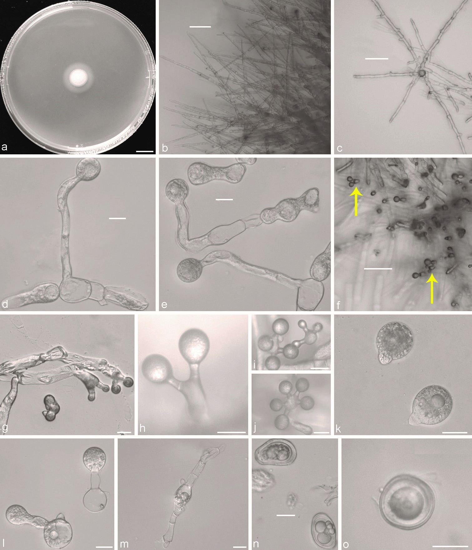

Conidiobolus taihushanensis sp. nov. a colony on PDA after 3 d at 21 °C b mycelia unbranched at the colony edge c young mycelia d, e primary conidiophores arising from mycelia segments f two branches germinated from hyphal bodies and each bearing a primary conidium (arrows) g– j two, three, four or five branches germinated from hyphal bodies and each bearing a primary conidium k globose to subglobose primary conidia l secondary conidia arising from primary conidia m zygospores formed between adjacent segments of the same hypha n young zygospores o mature zygospores. Scale bars: 10 mm(a); 100 μm (b, c, f); 20 μm (d, e, g–o).


