Conidiobolus bifurcatus B. Huang & Y. Nie, sp. nov. 2020
MycoBank No: 831599; Facesoffungi: FoF 08142
Holotype: China, Jiangsu: Nanjing, Laoshan National Forest Park, 32°6'7"N, 118°36'17"E, from plant debris, 1 Dec 2018, Y. Nie and Y. Gao (holotype HMAS 248359, ex-holotype culture CGMCC 3.15889 = RCEF 6551, GenBank: nucLSU = MN061285; TEF1 = MN061482; mtSSU = MN061288).
Morphological description
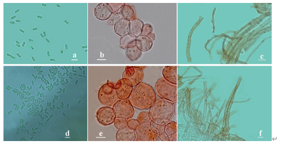
Colonies on PDA at 21 °C for 3 d, opaque, white, reaching ca. 2 mm in diameter, with many small colonies around the periphery due to discharged conidia. Mycelia colourless, 8–11 μm wide, rarely branched and non-septate when young, often septate and distended to a width of 10–27 μm after 5 d. Primary conidiophores arising from the hyphal segments, colourless, 38–254 × 7.5–12 μm, unbranched and producing a single globose conidium, without widening upwards near the tip. Primary conidia for- cibly discharged, globose to subglobose, 2–40 × 2–33 μm, with a papilla more or less tapering and pointed, 7–11 μm wide at the base, 3–12 μm long. Secondary conidiophores arising from the primary conidia, often branched almost at the tip, forming two short stipes each bearing a secondary conidium. Secondary conidia similar to, but smaller than the primary ones, mostly forcibly discharged, occasionally falling off and leaving a relic of the secondary conidiophores. On 2 % water agar, microconidia produced readily, globose to ellipsoidal, 7–12 × 6–9 μm. Zygospores homothallic, usually formed between adjacent segments of the same hypha after an incubation of 5–7 d at 21 °C on PDA, smooth, mostly globose, 25–40 μm in diameter, with a 1.5–3 μm thick wall.
Habitat: from plant debris
Distribution: Jiangsu Province, China.
GenBank Accession: nucLSU MN061285; EF-1α MN061482; mtSSU MN061288
Notes: Conidiobolus bifurcatus sp. nov. is characterised by its secondary conidiophores, which are often bifurcated near the tip and bear a secondary conidium on each stipe. Morphologically, it is allied to Conidiobolus mycophilus Srin. & Thirum., which has smaller primary conidia (Srinivasan and Thirumalachar 1965). It appears to be similar to C. incongruus Drechsler and C. mycophagus Srin. & Thirum. in the size of primary conidia and zygospores and the formation of microconidia, but different in its longer primary conidiophores (Drechsler 1960; Srinivasan and Thirumalachar 1965). However, it is distantly related to these two species in the molecular phylogenetic tree. Instead, it is phylogenetically closely related to C. brefeldianus Couch (Figure 1: MP 71/ML 89/BI 1.00), but morphologically distinct by its larger primary conidia and zygospores (Couch 1939).
Reference: Nie Y, Cai Y, Gao Y et al. (2020) Three new species of Conidiobolus sensu stricto from plant debris in eastern China.
Conidiobolus bifurcatus sp. nov. a Colony on PDA after 3 d at 21 °C b mycelium c septate mycelium and distended segments d, e primary conidiophores bearing primary conidia f, g primary conidia h, i a single secondary conidium produced from primary conidia j two secondary conidia arising from a branched conidiophore k secondary conidia falling from primary conidia l the relic of secondary conidiophores on secondary conidia (arrows) m microconidia arising from a conidium n, o globose microconidia p, q ellipsoidal microconidia r zygospores formed between adjacent segments of the same hypha s zygospores. Scale bars: 10 mm (a); 100 μm (b); 20 μm (c–s).


