 193
193
Cytospora spiraeicola H.Y. Zhu & X.L. Fan, sp. nov. 2020
MycoBank No 833821
Holotype: China, Beijing City, Mentougou District, Mount Dongling, Xiaolongmen Forestry Centre (115°28'28.52"E, 39°55'49.42"N), from branches of Spiraea salicifolia, 17 Aug 2017, H.Y. Zhu & X.L. Fan, holotype CF 2019803, ex-type living culture CFCC 53138.
Morphological description
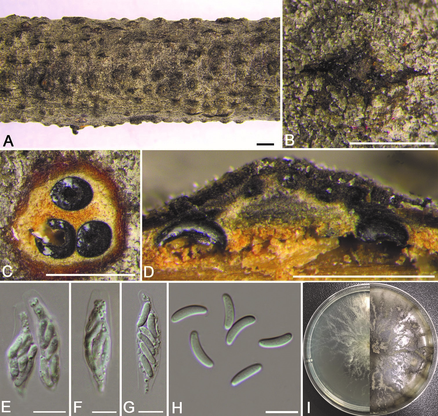
Necrotrophic on branches of Spiraea salicifolia and Tilia nobilis. Sexual morph: Ascostromata immersed in the bark, erumpent through the surface of bark, scattered, with 3–5 perithecia arranged regularly, 660–890 µm in diam. Conceptacle absent. Ectostromatic disc pale grey, usually surrounded by tightly crowded ostiolar necks, quadrangular, 240–350 µm in diam., with 5–8 ostioles arranged regularly per disc. Ostioles numerous, dark grey to black, at the same or above the level as the disc, concentrated, arranged regularly in a disc, 25–40 µm in diam. Perithecia dark grey to black, flask-shaped to spherical, arranged circularly, 210–250 µm in diam. Paraphyses lacking. Asci free, clavate to elongate, obovoid, 26–37 × 7.5–9 (av. = 33 ± 2.5 × 8.3 ± 0.9, n = 10) µm, 8-spored. Ascospores biseriate, elongate-allantoid, thin-walled, hyaline, slightly curved, aseptate, 8.5–12 × 2.5–3.5 (av. = 10 ± 1 × 3 ± 0.3, n = 30) µm. Asexual morph: not observed.Culture characteristics. Cultures are white, growing up to 4 cm in diam. with irregular margin after 3 days, covering the 9 cm Petri dish after 6 days, becoming vinaceous buff to hazel after 7–10 days. In reverse, the cultures are the same as the upper colour after 3 days, becoming isabelline to umber after 7–10 days. Colonies are felty with a heterogeneous texture, lacking aerial mycelium.
Habitat: from branches of Spiraea salicifolia
Distribution: Mount Dongling, China.
GenBank Accession: ITS MN85444; LSU MN85465,MN85466; act NA; rpb2 MN85074,MN85075; tef1-ΑMN85075; tub2 MN86111
Notes: Cytospora spiraeicola is associated with canker disease of Spiraea salicifolia and Tilia nobilis in China, with characteristics similar to Cytospora elaeagnicola and C. spiraeae in phylogram (Fig. 2). Morphologically, it differs from C. spiraeae by the smaller perithecia (210–250 vs. 270–400 µm in diam.) and longer ascospores (8.5– 12 × 2.5–3.5 vs. 7–8 × 2–2.5 µm) (Zhu et al. 2018a). Phylogenetically, C. spiraeicola (CFCC 53138) differs from C. elaeagnicola (CFCC 52882) by ITS (8/665), rpb2 (44/730), tef1-α (75/771) and tub2 (42/624) and C. spiraeae (CFCC 50049) by ITS (4/665), rpb2 (38/730), tef1-α (63/771) and tub2 (44/624) (Zhu et al. 2018a, Zhang et al. 2019). Therefore, we describe it as a novel species.
Reference: Zhu H, Pan M, Bezerra JDP et al. (2020) Discovery of Cytospora species associated with canker disease of tree hosts from Mount Dongling of China.
Cytospora spiraeicola from Spiraea salicifolia (CF 2019803). A, B habit of ascomata on twig C transverse section of ascoma D longitudinal section through ascoma E asci and ascospores F, G ascus H ascospores I colonies on PDA at 3 days (left) and 30 days (right). Scale bars: 1 mm (A, B); 500 µm (C, D); 10 µm (E–H).

