Inocybe squarrosofulva S.N. Li, Y.G. Fan & Z.H. Chen, sp. nov. 2021
MycoBank No: 839726
Holotype: China. Hunan Province: Zhangjiajie, Badagongshan National Nature Reserve, 29°67.57'N, 109°74.45'E, alt. 1600 m, on ground in subtropical montane forest, 29 July 2019, Z.H. Chen and S.N. Li, MHHNU31548 (GenBank accession no. ITS: MZ050799; nrLSU: MW715814; rpb2: MW574997).
Morphological description
Sexual morph:
Basidiomata. Small to medium-sized. Pileus 25–55 mm in diameter, spherical to bell-shaped when young, and gradually flattened to hemispheric or convex; margin strongly in-rolled when young then decurved or slightly uplifted; yellowish, center covered with yellow ochre to brownish yellow erect conical fibril lose scales (up to 1.5 mm high, 1–1.5 mm wide), coarsely fibrillose-rimose towards the margin; pileus with crenellated, nonpersisting fibrillose veil remnants at margin. Lamellae adnexed, crowded (ca. 55–70), up to 4 mm wide; yellowish brown, becoming brownish with age, edge concolorous. Stipe 40–80 × 5–8 mm, cylin drical, equal or slightly enlarged at the base, solid; light yellow to yellow ochre; pruinose with few yellowish-brown furfuraceous scales at apex; towards the base covered with numerous, yellow-ochre, woolly-fibrillose, incomplete zones; dry. Cortina conspicuous, annulate, composed of yellow ochre fibrils, and remains at the upper part of the stipe. Context: pale yellow in pileus and stipe. Odor like raw potatoes.
Basidiospores. (4.5) 5.0–7.0 μm (av. 6.6 μm, SD 1.0 μm) × 4.0–6.0 (7.0) (av. 5.3 μm, SD 0.8 μm) μm, Q = (1.00) 1.10–1.67 (1.75), Qm = 1.26 ± 0.16 (n = 80 of 4 coll.), nodulose with six hemispheric knobs, yellowish-brown with 5% KOH, containing a bright yellow oil droplet of uniform size inside. Basidia: 18–24 × 8–10 μm, 4-spored, clavate to broadly clavate. Pleurocystidia: 36–49 μm (av. 43.8 μm, SD 3.9 μm) × 13–18 μm (av. 15.5 μm, SD 2.6 μm), Q = 2.12–3.46 (n = 30 of 2 coll.), mostly hyaline, few with bright yellow oily inclusions, fusiform to broad ly fusiform, with crystalliferous apices, obtuse or truncated at base; thick-walled, walls bright yellow with 5% KOH, up to 2 μm thick towards apex. Cheilocystidia: 30–48 × 9–19 μm, similar to pleurocystidia, hyaline. Cheiloparacystidia: 10–23 × 6–12 μm, abundant among cheilocystidia, obovate, elliptic to clavate, thin-walled, hyaline. Hymenophoral trama: regular to subregular, composed of inflated hyphae, up to 18 μm wide, hyaline to lightly yellow with 5% KOH, thin-walled. Pileipellis: a trichoderm, subregular, consisting of cylindrical hyphae 5–13 μm in diameter, walls pale yellow brown with 5% KOH, smooth, thin-walled. Caulocystidia: pre sent at stipe apex, 23–49 × 9–21 μm, in clusters, thick-walled, walls thinner than pleurocystidia, hyaline or with pale yellow intracellular contents. Cauloparacystidia: 8–19 × 3–10 μm, clavate or broadly clavate, hyaline, thin-walled. Oleiferous hyphae present in pileus and stipe trama, 4–11 μm in diameter, branched. Clamp connec tions seen on all hyphae.
Asexual morph: Undetermined
Culture characteristics:
Habitat: On soil in subtropical montane forest dominated by Fagus lucida.
Distribution: Known from the type locality.
GenBank Accession:
ITS MZ050799; nrLSU MW715814; rpb2 MW574997
ITS MZ050802; nrLSU MW715815; rpb2 MW729766
Notes:
Reference: [1] Li, S. N. , Xu, F. , Jiang, M. , Liu, F. , & Chen, Z. H. . (2021). Two new toxic yellow inocybe species from china: morphological characteristics, phylogenetic analyses and toxin detection. MycoKeys, 81(1), 185-204.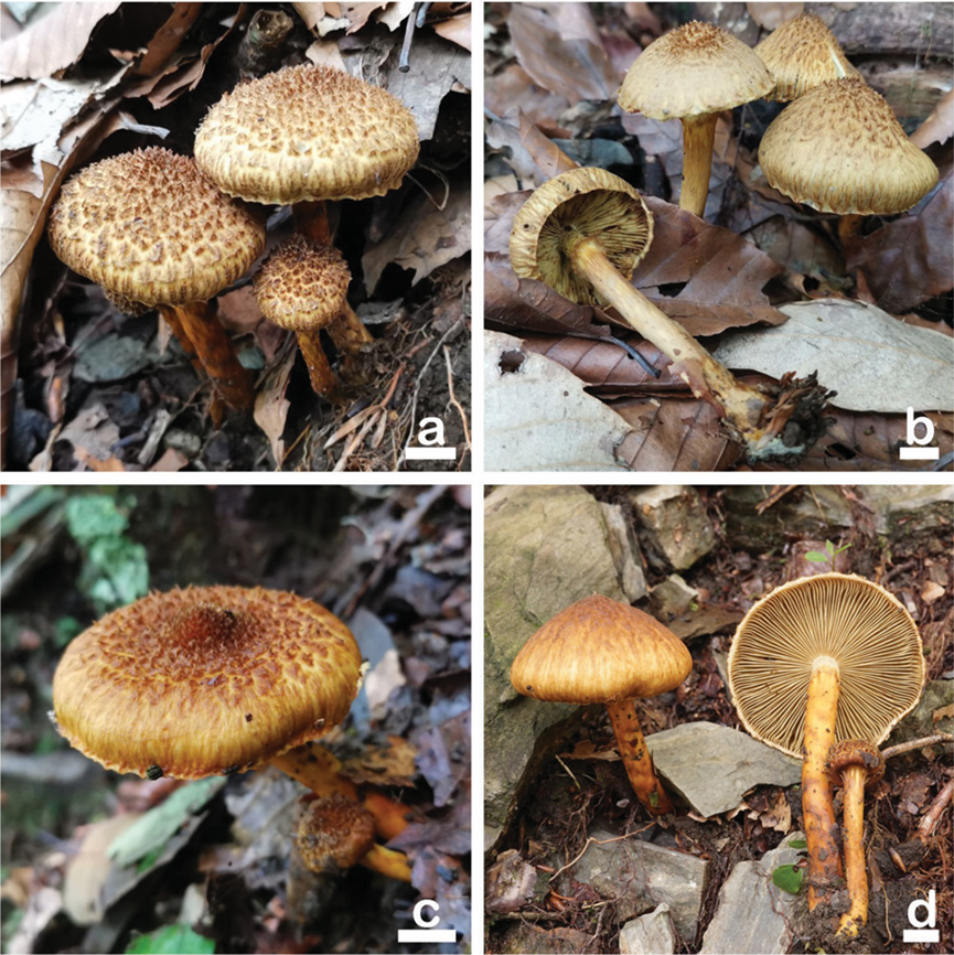
Figure 4. Basidiomata of Inocybe squarrosofulva a, b MHHNU31548 c, d MHHNU31927. Scale bars: 10 mm.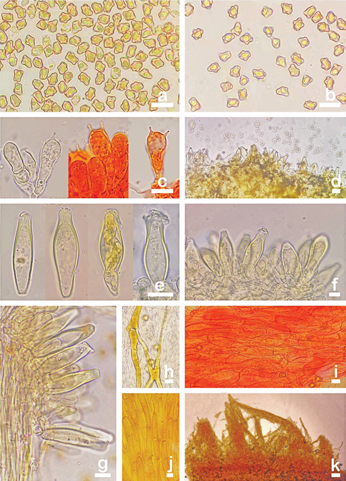
Figure 5. Microscopic features of Inocybe squarrosofulva (MHHNU31548, holotype) a, b basidiospores, c basidia with probasidium d gill edge e pleurocystidia f cheilocystidia and paracystidia g caulocystidia and cauloparacystidia h oleiferous hyphae i hymenial hyphae, and j, k pileipellis. Scale bars: 10 μm.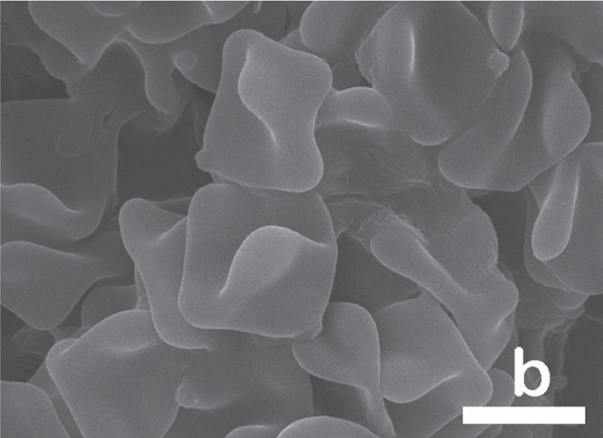
Figure 6. SEM images showing basidiospores of b Inocybe squarrosofulva. Scale bars: 5 μm.


