Atheniella rutila Q. Na & Y.P. Ge, sp. nov. 2021
MycoBank No: 839379
Holotype: China. Yunnan Province, Lincang City, Wulaoshan National Forest Park, 31 Jul 2020, Qin Na, Yupeng Ge and Zewei Liu, FFAAS0354 (Collection No. MY0210).
Morphological description
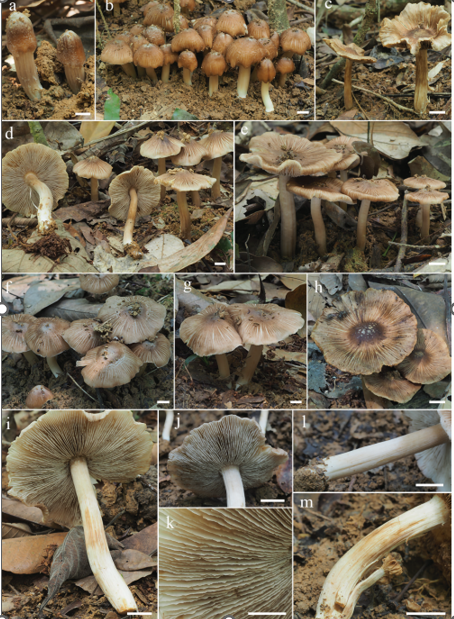
Sexual morph: Pileus 2.0–10.2 mm in diam., campanulate to hemispherical, ap planate or slightly concave at centre when old, deep salmon to bright red, shallowly sulcate, translucent-striate, delicately pubescent, glabrescent when old. Context white, thin, very fragile. Lamellae broadly adnate to adnexed, ascend ing, white, concolorous with the sides, basally interveined with age. Stipe 5.0–15.8 × 1.0–2.0 mm, cylindrical, hollow, fragile, transparent, pruninose, glabrescent when old, base slightly swollen, covered with dense white fibrils. Odour and taste indistinctive.
Basidiospores [60/3/2] (7.2) 7.7–8.6–9.8 (10.1) × (3.6) 4.1–4.6–5.3 (5.5) μm [Q = 1.71–2.05, Q = 1.85 ± 0.079] [holotype [40/2/1] (7.2) 7.5–8.5–9.7 (10.0) × (3.6) 4.1–4.6–5.2 (5.5) μm, Q = 1.72–1.99, Q = 1.86 ± 0.086], narrowly ellipsoid to cylin drical, hyaline in water and 5% KOH, inamyloid, smooth. Basidia 19–28 × 5–8 μm, 2-spored, clavate, hyaline. Cheilocystidia 32–45 × 8–11 μm, abundant, fusiform, with a long neck, thin-walled and hyaline. Pleurocystidia similar to cheilocystidia, 27–42 × 7–12 μm. Pileipellis hyphae 2–5 μm wide, covered with numerous excrescences, 3.2 6.9 × 0.8–1.7 μm, hyaline. Hyphae of the stipitipellis 2–7 μm wide, non-dextrinoid, hyaline, with simple cylindrical excrescences, 4.6–14.3 × 2.9–5.2 μm. All tissues non reactive in iodine. Clamps absent in all tissues.
Asexual morph: Undetermined
Culture characteristics:
Habitat: Scattered on rotten wood in evergreen broadleaf and Pinus mixed forest.
Distribution: China
GenBank Accession:
ITS Sequences ID MW969658; nLSU Sequences ID MW969668
ITS Sequences ID MW969659; nLSU Sequences ID –
Remarks: Atheniella rutila is considered to be a distinct species in Atheniella on ac count of the bright red pileus, white stipe, narrowly ellipsoid to cylindrical and inamy loid spores and characters of the cystidia, pileipellis and stipitipellis (Maas Geesteranus 1980, 1990, 1992a, b; Perry 2002; Grgurinovic 2003; Robich 2003; Aravindakshan and Manimohan 2015; Aronsen and Læssøe 2016). Atheniella amabillissima is diffi cult to distinguish from A. rutila owing to the reddish basidiomata, but the pileus of A. amabillissima fades to white with age or has a dirty yellowish disc, and the spores are smaller (7–9 × 3–4 μm) (Smith 1935b). Atheniella adonis shows certain morphological similarities to A. rutila in possessing tiny and pinkish-red basidiomata, white lamellae and cylindrical basidiospores. However, A. adonis differs in producing a pileus with a white margin, longer stipe and clavate to fusiform caulocystidia (Perry 2002; Robich 2003; Aronsen and Læssøe 2016). In comparison with Atheniella rutila, Mycena rohitha (≡ A. rohitha) and M. wubabulna (≡ A. wubabulna) have gelatinous pileus hyphae and narrower basidiospores (Grgurinovic 2003; Aravindakshan and Manimohan 2015).
Reference: [1] Ge, Y. , Liu, Z. , Zeng, H. , Cheng, X. , & Na, Q. . (2021). Updated description ofatheniella(mycenaceae, agaricales), including three new species with brightly coloured pilei from yunnan province, southwest china. MycoKeys, 81, 139-164.
Figure 2. Basidiomata of Atheniella species m–p Atheniella rutila Q. Na & Y.P. Ge. Scale bars: 10 mm (n-p).
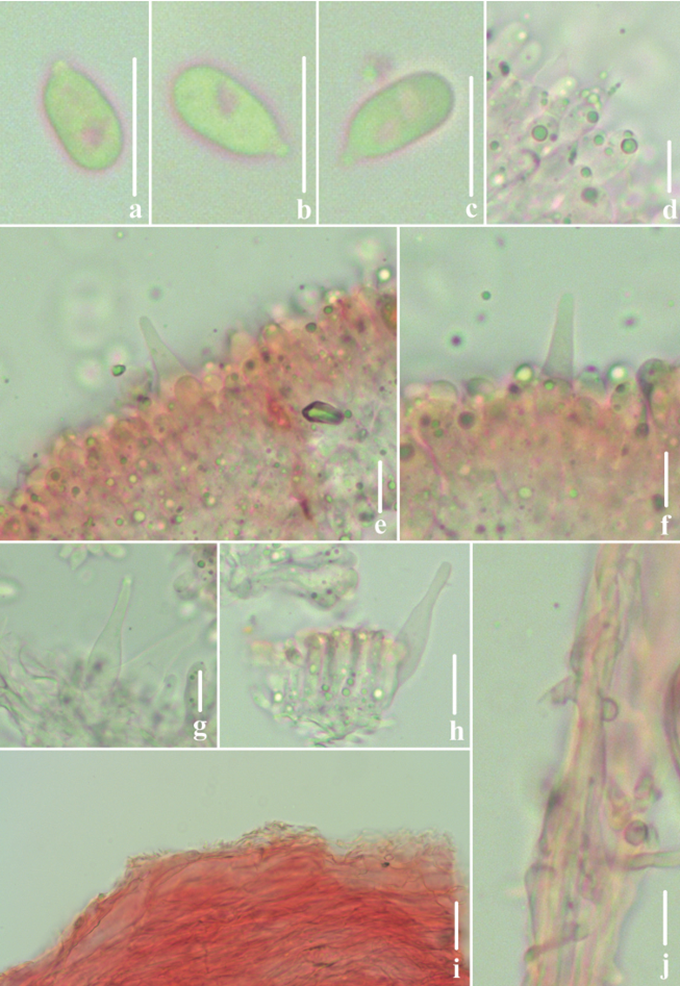
Figure 5. Microscopic features of Atheniella rutila (FFAAS0354, holotype) a–c basidiospores d basidia e, f cheilocystidia g, h pleurocystidia i pileipellis j stipitipellis. Scale bars: 10 μm (a–j). 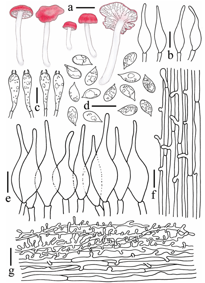
Figure 6. Morphological features of Atheniella rutila (FFAAS0354, holotype) a basidiomata b pleuro cystidia c basidia d basidiospores e cheilocystidia f stipitipellis g pileipellis. Scale bars: 5 mm (a); 10 μm (b–g). Drawings by Qin Na and Yupeng Ge.


