 83
83
Diaporthe hunanensis Q. Yang, sp. nov.2021
MycoBank No: 840452
Holotype:
Morphological description
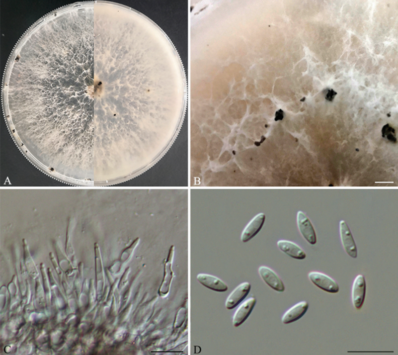
Asexual morph: pycnidia on PDA 180–300 μm in diam., superficial, scattered, black, globose, solitary in most. Conidiophores reduced to conidiogenous cells. Conidiogenous cells (8–)9–15(–16.5) × 1.7–2.1 μm (n = 30), aseptate, cylindrical, phiailidic, straight or slightly curved. Alpha conidia 6.5–7.5(–8.5) × 2.4–2.9 μm (n = 30), aseptate, hyaline, ellipsoidal, biguttulate, both ends obtuse. Beta conidia not observed.
Sexual morph: Undetermined
Cultures: Culture incubated on PDA at 25 °C, originally flat with white fluffy aerial mycelium, becoming pale brown with age, with visible solitary conidiomata at maturity after 18 days.
Habitat: on leaves of Camellia oleifera,
Distribution: . China. Hunan Province: Zhuzhou City
GenBank Accession: : HNZZ025 and HNZZ033).
Notes: Three isolates representing D. hunanensis cluster in a well-supported clade (ML/BI=100/1) and appear most closely related to D. chrysalidocarpi on Chrysalidocarpus lutescens, D. drenthii and D. searlei on Macadamia sp., and D. spinosa on P. pyrifolia cv. Cuiguan. Diaporthe hunanensis can be distinguished from D. chrysalidocarpi based on ITS, cal, his3 and tub2 loci (7/457 in ITS, 28/448 in cal, 8/455 in his3 and 5/401 in tub2); from D. drenthii based on ITS, tef1 and tub2 loci (9/457 in ITS, 13/328 in tef1 and 23/401 in tub2); from D. searlei based on ITS and tub2 loci (10/457 in ITS and 12/401 in tub2); from D. spinosa based on ITS, cal, his3, tef1 and tub2 loci (8/458 in ITS, 31/448 in cal, 5/455 in his3, 8/328 in tef1 and 19/401 in tub2). Morphologically, D. chrysalidocarpi produces only beta conidia, while D. hunanensis produces alpha conidia (Huang et al. 2021); D. hunanensis differs from D. drenthii and D. searlei in wider alpha conidia (2.4–2.9 μm in D. hunanensis vs. 1.5–2.5 μm in D. drenthii vs. 1.5–2 μm in D. searlei) (Wrona et al. 2020); from D. spinosa in shorter alpha conidia (6.5–7.5 × 2.4–2.9 μm vs. 5.5–8 × 2–3.5 μm) (Guo et al. 2020). Therefore, we establish this fungus as a novel species.
Reference:[1] Mi, L. I. , Zhen, H. E. , Xiaowu, L. , Yonggang, X. , Xuewu, Y. , & Academy, H. F. . (2015). Investigation on resources and appraisal of dominant species of natural enemies in camellia oleifera forests in hunan province. Hunan Forestry Science & Technology.

Diaporthe hunanensis (HNZZ023) A Culture on PDA B conidiomata C conidiogenous cells D alpha conidia. Scale bars: 500 μm (B); 10 μm (C–D).

