Colletotrichum xishuangbannaenseN.I. de Silva & Lumyong, sp. nov. 2021
MycoBank Index Fungorum number: IF556380; Facesoffungi number: FoF09495
Holotype: MFLU 20–0593
Morphological description
Sexual morph:
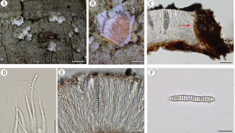
Asexual morphs: coelomycetous, conidial masses with mycelia in culture on PDA. Conidiophores not observed. Conidiogenous cells 15–18 × 1.5–2 µm (x= 16 × 1.7 µm), hyaline, cylindrical, smoothwalled. Conidia 9–12 × 3–4 µm (x= 11 × 3.5 µm), hyaline, cylindrical with the apex and base rounded. Appressoria not observed.
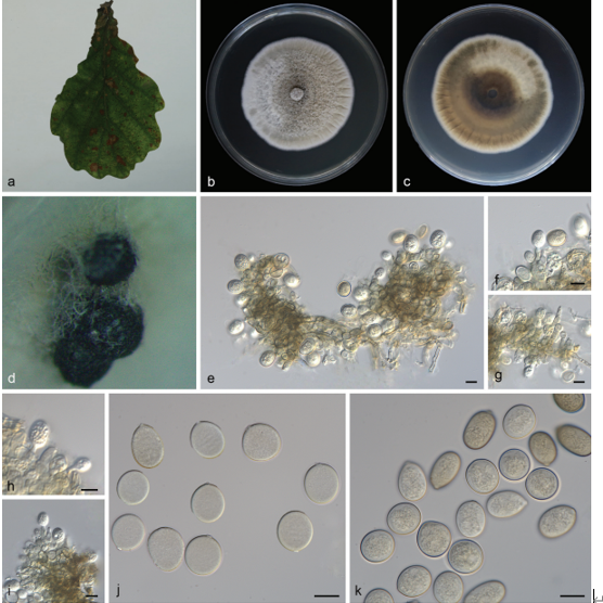
Cultures: Colonies on PDA reaching 27 mm diameter after 7 days at 25°C, colonies circular, margin undulate, slightly raised, velvety appearance, cream aerial mycelia and colony from above: light brown; reverse: brown.
Habitat: healthy leaves of Magnolia candolli (Magnoliaceae)
Distribution: CHINA, Yunnan Province, Xishuangbanna.
GenBank Accession: (S17); ITS: MW346469, GAPDH: MW537586, CHS: MW660832, ACT: MW652294, (S118); ITS: MW346470, GAPDH: MW537587, CHS: MW660833, ACT: MW652295.
Notes: Colletotrichum xishuangbannaense clustered closely related to C. aeschynomenes with 40% MP, 25% ML, 0.83 BYPP support (Fig. 19). Morphologically, C. xishuangbannaense differs from C. aeschynomenes in having small conidia. Colletotrichum aeschynomenes has 14–20 × 4–5 µm conidia (Weir et al. 2012) and C. xishuangbannaense has 9–12 × 3–4 µm conidia. Colletotrichum magnoliae was isolated from leaves of Magnolia grandiflora in Portugal (Saccardo 1931). Conidia of C. magnoliae (15−18 × 5−6 µm) (Saccardo 1931) are larger than C.xishuangbannaense.
Reference: de Silva NI, Maharachchikumbura SSN, Thambugala KM, Bhat DJ, Karunarathna SC, Tennakoon DS, Phookamsak R, Jayawardena RS, Lumyong S, Hyde KD 2021 – Morphomolecular taxonomic studies reveal a high number of endophytic fungi from Magnolia candolli and M. garrettii in China and Thailand. Mycosphere 11(1), 163–237, Doi 10.5943/mycosphere/12/1/3
Colletotrichum xishuangbannaense (MFLU 20–0593, holotype). a Colonies on PDA. b Conidial masses. c Mycelia and conidial masses. d Conidiogenus cells with conidia. e, f Conidia. Scale bars: c = 50 μm, d–f = 10 μm.