Coryneum suttonii C.M. Tian, Voglmayr & N. Jiang 2020
MycoBank MB824596.
Holotype: CHINA. SHAANXI PROVINCE: Shangluo City, chestnut plantation, 33°39′27.25″N, 109°07′15.48″E, 2504 m asl, on branches of Castanea mollissima, N. Jiang, 8 Jul 2017 (holotype BJFC-S1423). Ex-type culture: CFCC 52317.
Morphological description
Sexual morph: Not observed.
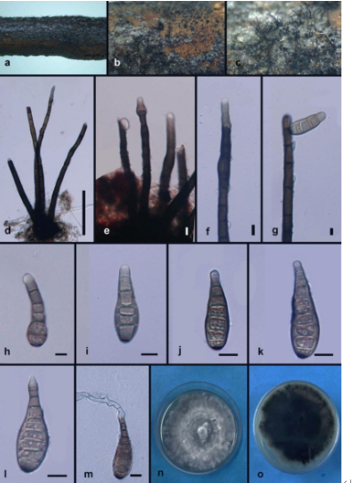
Asexual morph: Conidiomata acervular, 0.5–2.0 mm wide, 0.3–1.2 mm high (x = 1.0 × 0.6 mm, n = 20), solitary, erumpent through outer periderm layers of host, scattered, surface tissues above slightly domed. Conidiophores 40–90 μm long, 4–8 μm wide (x = 70× 6 μm, n = 20), unbranched, cylindrical, septate, hyaline at apex, pale brown at the base. Conidiogenous cells holoblastic, integrated, indeterminate, cylindrical, expanding toward apices, hyaline, smooth, with 0–1 percurrent extensions. Conidia (60–)68–74(–76) × (10–)10.5–13(–14.5) μm, L/W = (4.8–)6.4–6.5(–6.6) (n = 50), curved or not, fusiform to clavate, dark brown, smooth-walled, 4–5-distoseptate, apical cell with a hyaline tip, truncate and black at base.
Culture characters: On PDA at 25 C, colonies growing slowly and unevenly, reaching 60 mm diam within 30 d, becoming brownish gray to dark gray in color with scant cottony aerial mycelium, asexual morphs developed after 2 mo.
Habitat: On branches of Castanea mollissima.
Distribution: In China.
GenBank Accession: ITS : MH683555;28S: MH683563; TEF1α: MH685735; RPB2: MH685727
Notes: With the addition of two new species in the present publication, four Coryneum species now are known from chestnut trees (Castanea spp.). Coryneum
suttonii can be distinguished from C. gigasporum by smaller conidia (60–76 × 10–14.5 μm in C. suttonii vs. 88–117 × 18–23 μm in C. gigasporum), and from C. castaneicola by fewer distosepta (4–5 in C. suttonii vs. 6–7 in C. castaneicola). The conidial length of C. suttonii is similar to that of C. modonium (60–76 μm in C. suttonii vs. 50–71 μm in C. modonium), but conidia of C. suttonii are distinctly narrower (10–14.5 μm in C. suttonii vs. 14–19 μm in C. modonium).
Reference: Ning Jiang a, Hermann Voglmayr b, and Chengming Tian a

Morphology of Coryneum suttonii from Castanea mollissima (BJFC-S1423, holotype). A, B. Conidiomata on natural substrate in surface view. C. Transverse section through conidioma. D. Longitudinal section through conidioma. E, F. Conidiophores. G–J. conidia. Bars: A–D = 0.5 mm; E–J = 10 μm.