Tyromyces minutulus Y.C. Dai & C.L. Zhao 2020
Index Fungorum number: IF556446; Facesoffungi number: FoF 06044
Holotype: CHINA, Hainan Province, Changjiang County, Bawangling Nature Reserve, on rotten angiosperm wood, 16 June 2014 Dai 13666 (holotype); on fallen angiosperm trunk, 8 May 2009 Cui 6427 (BJFC).
Morphological description
Basidiocarp annual, pileate, occasionally attached by a lateral tapering base, soft and sappy when fresh, becoming corky upon drying. Pilei more or less semicircular, projecting up to 1 cm, 1.5 cm wide, 1.5 mm thick at base. Pileal surface concentrically zonate, white when juvenile, becoming light chocolate to pale brown in centre, more or less glabrous to slightly rough. Pore surface white when fresh, white to cream upon drying; pores round to angular, 7–9 per mm; dissepiments thin, entire. Sterile margin white, up to 2 mm wide. Context white, corky, up to 0.5 mm thick. Tubes concolorous with pore surface, corky, up to 1 mm long. Hyphal system monomitic; generative hyphae with clamp connections, IKI–, CB–; tissues unchanged in KOH. Subicular generative hyphae hyaline, thick-walled with a wide lumen, unbranched, interwoven, 2.5–4.5 µm diam. Tramal generative hyphae hyaline, thin- to thick-walled, frequently branched, interwoven, 2.0–3.0 µm diam. Cystidia absent, but fusoid cystidioles present, hyaline, thinwalled, 7.0–8.0 × 3.0–3.5 µm; basidia more or less barrelshaped, with four sterigmata and a basal clamp connection, 9.0–11.0 × 3.0–4.5 µm; basidioles dominant; basidioles in shape similar to basidia, but slightly smaller. Basidiospores cylindrical, hyaline, thin-walled, smooth, tapering at apiculus, usually bearing one or two guttules, IKI–, CB–, (3.5–) 3.7–4(–4.2) × 1.0–1.3(–1.5) µm, L = 3.89 µm, W = 1.18 µm, Q = 3. 19-3.45 (n = 60/2).
Habitat: On rotten angiosperm wood.
Distribution: In China.
GenBank Accession: ITS: KM598443; LSU: KM598445.
Notes: Phylogenetically, the new species, Tyromyces minutulus, groups with T. chioneus, T. kmetii (Bres.) Bondartsev & Singer, Piloporia sajanensis and Skeletocutis amorpha (Fig. 107). However, morphologically T. chioneus differs from T. minutulus by its larger pores (3–4 per mm) and basidiospores (4.0–5.0 × 1.5–2.0 µm, Núñez and Ryvarden 2001). T. kmetii is separated from T. minutulus by the orange pore surface and larger basidiospores (4.0–4.5 2.5–3.0 µm, Gilbertson and Ryvarden 1987). Skeletocutis amorpha differs in its resupinate to effuso-reflexed basidiocarps, a dimitic hyphal system and allantoid basidiospores (Núñez and Ryvarden 2001; Ryvarden and Melo 2014). Piloporia sajanensis is distinguished from T. minutulus by the distinctly duplex structure of the context and the brown hyphae (Niemelä 1982). Morphologically the basidiospores of the new species are reminiscent of several similar Tyromyces species. T. polyporoides Ryvarden & Iturriaga differs in its laterally stipitate basidiocarps (Ryvarden and Iturriaga 2003); T. americanus (D. Reid) Ryvarden & Iturriaga differs from T. minutulus by substipitate basidiocarps and pale orange pore surface (Ryvarden and Iturriaga 2003); T. leucomallus (Berk. & M.A. Curtis) Murrill differs in having larger basidiocarps and gloeoplerous hyphae (Núñez and Ryvarden 2001); and T. subviridis Ryvarden & Guzmán is distinguished by ochraceous basidiocarps and a grey to pale green pore surface (Ryvarden and Guzmán 2001).
Reference: Hai‑Sheng Yuan1,2· Xu Lu1,2 · Yu‑Cheng Dai3 ·
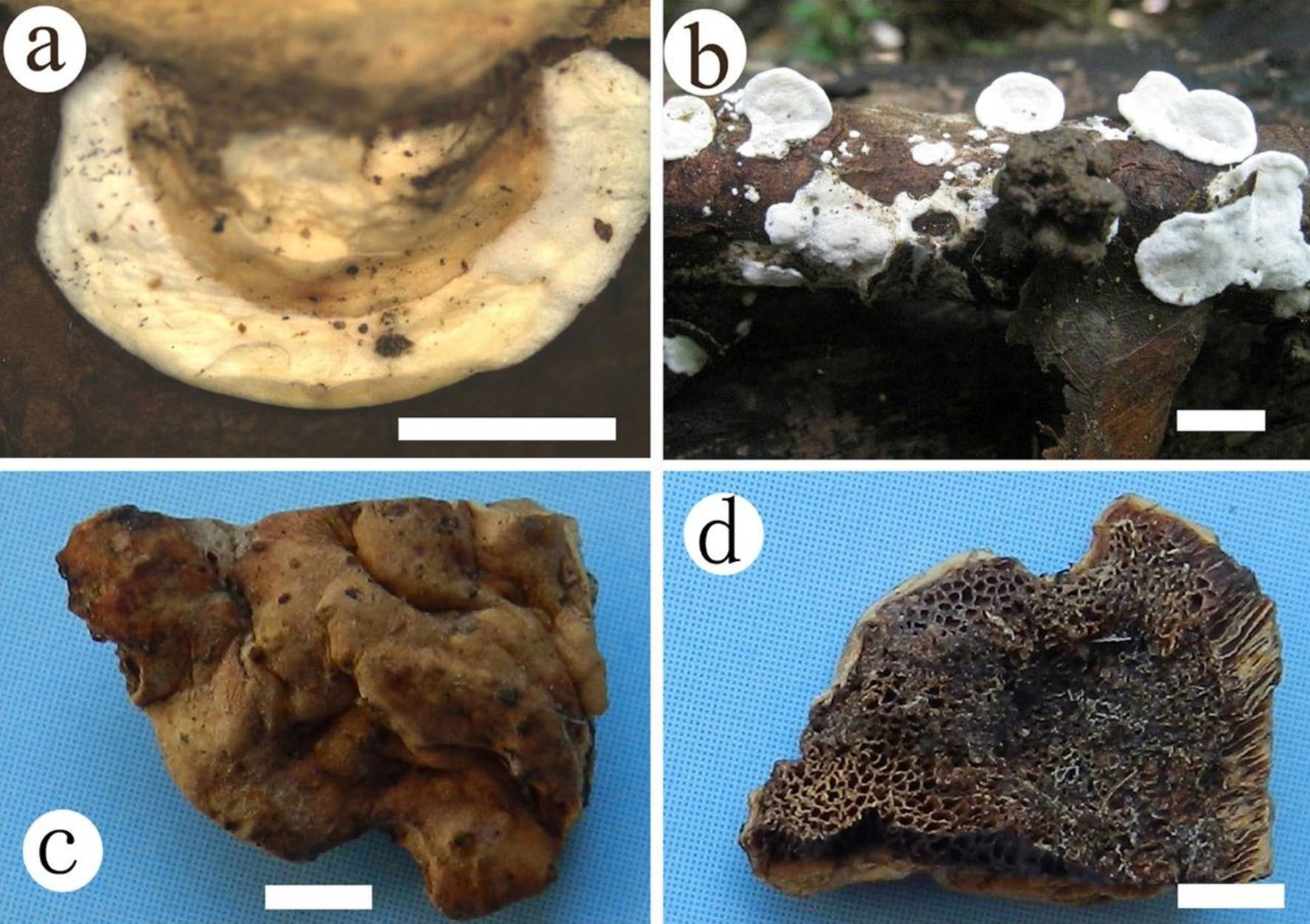
Basidiocarps of Tyromyces minutulus (BJFC017406, holotype) and the combination taxon Antrodiella descendena (holotype). a, b Tyromyces minutulus. c, d Antrodiella descendena. Scale bars: a = 0.5 cm, e–f = 2 mm