Amphisphaeria camelliae Samarak., Jian K. Liu & K.D. Hyde, sp. nov. 2020
MycoBank: MB836110; Facesoffungi number: FoF08740
Holotype: China, Sichuan Province, Chengdu, University of Electronic Science and Technology of China (UESTC) campus, on the wood of Camellia japonica (Theaceae), 30 September 2019, M.C. Samarakoon, SAMC254 (HKAS 107021, holotype; MFLU 20-0504, isotype); ex-type living culture MFLUCC 20-0122.
Morphological description
Saprobic on the dead wood of Camellia japonica (Theaceae). Sexual morph: Ascomata: 300–480 µm high × 160–310 µm diameter (M = 410 × 260 µm, n = 5), immersed, visible as black spots covered with pale brown and blackish area, solitary or aggregated, scattered, globose to subglobose, papillate ostiole 80–150 µm high × 50–85 µm diameter (M = 110 × 60 µm, n = 5), centric. Periphyses: 1–2 µm wide (M = 1.5 µm, n = 20), hyaline, short. Peridium: two-layered; outer layer 5–7 µm (M = 5.5 µm, n = 10), dense, reddish-brown cells of textura angularis 8–15 × 1–2 µm (M = 10.8 × 1.6 µm, n = 15), thick-walled. Inner layer: 7–12 µm (M = 9.5 µm, n =10), loosely arranged, hyaline cells of textura angularis 9.5–18 × 1–3 µm (M = 12 × 1.5 µm, n = 15), thin-walled, loosely arranged. Paraphyses: 2–4 µm wide (M = 3 µm, n = 20), hyaline, highly delicate, cellular, constricted septate, guttulate; 1–2 µm wide (M = 1.5 µm, n = 20), hyaline, filiform, longer than asci, blunt end, cellular, guttulate, embedded in a gelatinous matrix. Asci: 85–130 × 5–8 µm (M = 110 × 6.5 µm, n = 25), 8-spored, unitunicate, cylindrical, thin-walled, short-pedunculate, apically rounded, with a J+, discoid apical ring. Ascospores: 12–17.5 × 4–5.5 µm (M = 15 × 5 µm, n = 40), l/w 3.1, uniseriate, oblong or narrowly fusiform, first hyaline, guttulate, turning yellow to yellowish-brown, with a median septum, slightly constricted at the septum, straight to slightly curved, smooth-walled. Asexual morph: Coelomycetous. Conidiomata: superficial on PDA, solitary or aggregated, globose, dark brown. Conidiophores: 24–40 × 1–3 µm (M = 31 × 2 µm, n = 15), arising from peridium, septate, branched, thick-walled, light brown to hyaline. Setae: 60–92 × 3.5–5 µm (M = 76 × 4.5 µm, n = 5), septate, thick-walled, blunt end, brown to light brown. Conidiogenous cells: 7.5–14.5 ×1.5–2.5 µm (M = 11.5 × 2 µm, n = 15), elongated conical, thin-walled, hyaline, annellidic, guttulate. Conidia: 14.5–18 × 1.5–2.5 µm (M = 16 × 2 µm, n = 25), elongate-fusiform, curved, smooth-walled, hyaline, guttulate. Culture characteristics: colonies on PDA, reaching 16–17 mm diameter after one week at 25 ◦C, the colonies are flat, circular, dense, with a smooth surface, entire margin, and white to light brown.
Media become pale brown; reverse light brown at center and dirty white edges.
Habitat: on the wood of Camellia japonica (Theaceae).
Distribution: Sichuan Province, Chengdu, China.
GenBank Accession: LSU MT756615; ITS MT756621; RPB2 MT789850; TUB2 MT774368
Notes: Our specimens have solitary and aggregated, immersed ascomata with two-layered peridium, unitunicate asci with J+, a discoid apical ring, and brown ascospores similar to amphisphaeriaceous species. Amphisphaeria camelliae possesses 1-septate ascospores similar to A. uniseptata, but differs in having single or aggregated, globose to subglobose (vs. single, subglobose or applanate) ascomata, thin paraphyses (3.1 vs. 5 µm) and large ascospores (l/w 3.1 vs. l/w 2.7). In the phylogenetic analyses, our collection also clusters with A. uniseptata. Based on morphology and phylogeny, our collection is introduced as a novel species A. camelliae.
Reference: Milan C. Samarakoon 1,2,3,4,5 ,Sajeewa S. N. Maharachchikumbura,Jian-Kui (Jack) Liu 4 et al.
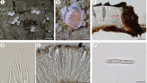
The sexual morph of Amphisphaeria camelliae (HKAS 107021, holotype). (a,b) Host Camellia japonica; (c) substrate; (d–f) ascomata on the substrate; (g) vertical section of ascoma; (h) peridium; (i) ostiole; (j) paraphyses; (k–n) asci; (o,p) apical ring bluing in Melzer’s reagent; (q) ascospore top view; (r–w) ascospores (v in Congo Red). Scale bars are set at (c) 1 cm; (f) 500 µm; (d,e) 200 µm; (g) 100 µm; (h,i,k–n) 20 µm; (o,p,r–w) 10 µm; (q) 5 µm.
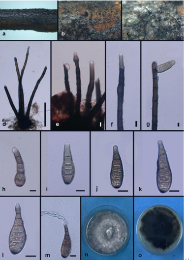
The asexual morph of Amphisphaeria camelliae (MFLUCC 20-0122, ex-type living culture).
(a) Germinating ascospore; (b) upper view, (c) reverse view of the 2 weeks old colony on PDA; (d) conidiomata in the culture; (e) setae; (f–h) conidiophores, conidiogenous cells and conidiogenesis; (i–n) conidia. Scale bars are set at (e) 50 µm; (a,f–h) 10 µm; (i–n) 5 µm.