Dictyosporium muriformis N.G. Liu, K.D. Hyde & J.K. Liu 2020
Index Fungorum number: IF557096; Facesoffungi number: FoF 06707
Holotype: CHINA, Guizhou Province, Dushan, on decaying wood in a bank of a small freshwater, 6 July 2018, N.G. Liu, DS024 (MFLU 19-2853, holotype), ex-type living culture, GZCC 20-0006.
Morphological description
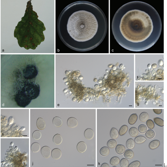
Sexual morph Undetermined. Asexual morph Hyphomycetous. Colonies on natural substrate sporodochial, scattered, black, glistening. Conidiophores semi-micronematous, mononematous, subhyaline to pale brown, aseptate, cylindrical, smooth-walled, thinwalled. Conidiogenous cells 3–3.7 μm wide, monoblastic, for LSU, 580 characters for ITS, 922 characters for TEF1-α) after alignment. Periconia igniaria (CBS 379.86) in Periconiaceae (Pleosporales) is used as the outgroup taxon. Single gene analyses were also performed to compare the topology and clade stability with combined gene analyses. Tree topology of the maximum likelihood analysis is similar to the Bayesian analysis. The best RaxML tree with a final likelihood values of − 13815.246477 is presented. The matrix had 844 distinct alignment patterns, with 31.91% undetermined characters or gaps. Estimated base frequencies were as follows: A = 0.234018, C = 0.251157, G = 0.271122, T = 0.243703; substitution rates AC = 1.698556, AG = 3.692510, AT = 2.823016, CG = 0.799026, CT = 9.194311, GT = 1.000000; gamma distribution shape parameter α = 0.186936. Bootstrap values for maximum likelihood (ML) equal to or greater than 75% and clade credibility values greater than 0.95 from Bayesian-inference analysis labeled on the nodes. The new isolates are indicated in bold and blue integrated, terminal, subhyaline to pale brown, enlarge at apex. Conidia 20–30 × 11–14.5 μm ( x̄ = 24.5 × 14.5 μm, n = 20), solitary, acrogenous, cheiroid, median brown, not complanate, guttulate, consisting of 14–27 cells arranged in (3–)4(–5) closely compact rows, 2–7-euseptate in each column.Culture characteristics: Conidia germinating on water agar within 24 h. Germ tubes produced from the base of conidia. Mycelia superficial, dense, circular, with entire edge, greyish brown from above and below.
Habitat: decaying wood
Distribution:China.
GenBank Accession: ITS:MT002304,;LSU:MN897834;SSU:MN901117;TEF1-α:MT023011;RPB2:MT023014.
Notes: Phylogenetic analyses (Fig. 17)show that Dictyosporium muriformis is basal to D. meiosporum Boonmee & K.D. Hyde, and D. tetrasporum L. Cai & K.D. Hyde and constitute an independent lineage within Dictyosporium. The conidia of D. muriformis resemble those of other species which consist mainly of four compact arms. However, D. nigroapice and D. tubulatum have conidial appendages which are absent in D. muriformis. Moreover, the conidia of D. muriformis are thinner than those of D. tetrasporum (11–14.5 versus 16–21.5 μm), and wider than those of D. meiosporum (11–14.5 versus 6–8.5 μm).
Reference: Kevin D. Hyde1,5,8,22 · Yang Dong2,3 · Rungtiwa Phookamsak1,5,6,7 et al.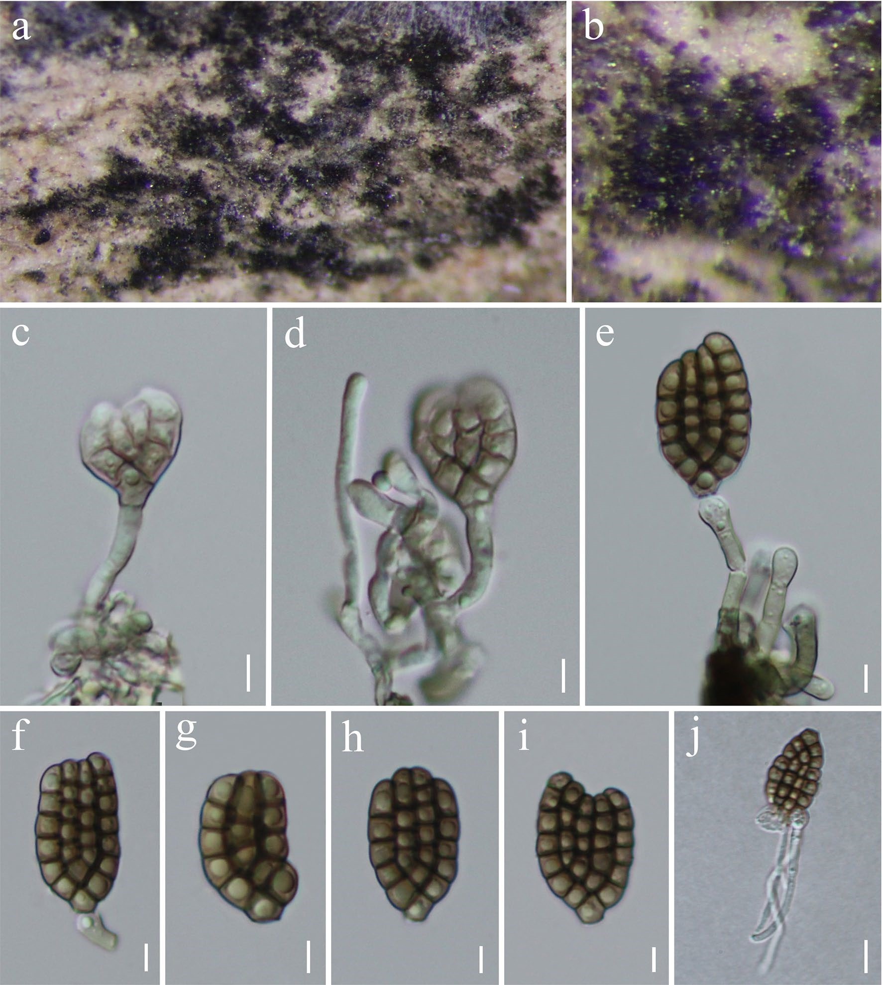
Dictyosporium muriformis (MFLU 19-2853, holotype). a, b Colonies in natural substrates. c–e Conidiophores, conidiogenous cells and conidia. f–i Conidia. j Germinated conidium. Scale bars: c–i = 5 μm, j = 10 μm